IMAGE IN CARDIOLOGY

DOI: 10.4244/EIJV11I4A88

Very late metallic stent malapposition and in-stent restenosis treated with a bioresorbable scaffold: a novel alternative for an old problem

A 62-year-old man had stenting of the proximal left anterior descending artery with a 3.5×23 mm CYPHER® drug-eluting stent (DES) (Cordis, Johnson & Johnson, Warren, NJ, USA) 10 years ago. He presented with unstable angina. Optical coherence tomography (OCT) revealed evidence of neoatherosclerosis and a minimal luminal area (MLA) of 2.08 mm2 as well as evagination with extensive late stent malapposition (Figure 1A-Figure 1C, Moving image 1).

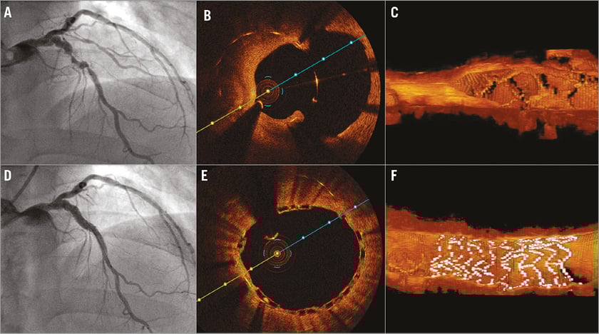
Figure 1. Very late metallic stent thrombosis and in-stent restenosis treated with a BVS. A) In-stent restenosis and focal aneurysm in the left anterior descending artery around the sirolimus DES implanted for 10 years. B) Neointimal proliferation and evagination with stent malapposition detected on 2D and 3D OCT (C). D) Final angiography and OCT (E & F) reveal the adequate scaffold expansion and apposition with an MLA of 9.74 mm2.

After re-dilatation of the lesion, drug-eluting balloons (DEB) were applied across the stented segment and a 3.5×12 mm Absorb Bioresorbable Vascular Scaffold (BVS) (Abbott Vascular, Santa Clara, CA, USA) was deployed proximally to cover the aneurysm and a proximal stent-edge dissection. This was followed by post-dilation of the scaffolded segment using a 4.0×15 mm NC balloon. Final angiography and OCT study revealed adequate scaffold expansion and apposition with an MLA of 9.74 mm2 (Figure 1D-Figure 1F).

OCT imaging can provide valuable insights into late failure of metallic DES. Late stent malapposition has been reported following DES implantation and is recognised as a hallmark for late stent thrombosis. Here the patient presented with both restenosis and aneurysm formation. Implantation of another DES may continue to aggravate the late stent malapposition issue. On the other hand, use of bare metal stents would result in a high likelihood of recurrent restenosis or neoatherosclerosis. Hence, BVS may be a reasonable alternative in the treatment of patients with such late DES failure.

Conflict of interest statement

The authors have no conflicts of interest to declare.

Online data supplement

Moving image 1. 3D OCT of the CYPHER evagination and late stent malapposition.

Supplementary data

To read the full content of this article, please download the PDF.

Moving image 1. 3D OCT of the CYPHER evagination and late stent malapposition.


Key metrics

Suggested by Cory

IMAGE IN CARDIOLOGY

10.4244/EIJV12I1A10 May 16, 2016
Coronary aneurysm without malapposition after bioresorbable vascular scaffold implantation
Timmers L et al
free

IMAGE IN CARDIOLOGY

10.4244/EIJ-D-16-00323 Jan 20, 2017
Delayed fracture of a bioresorbable vascular scaffold implanted for in-stent restenosis
Bastante T et al
free

Image – Interventional flashlight

10.4244/EIJ-D-19-00063 Jan 20, 2021
Late structural discontinuity after bioresorbable vascular scaffold implantation in patients with in-stent restenosis
Cuesta J et al
free

IMAGE IN CARDIOLOGY

10.4244/EIJY14M10_07 Nov 20, 2015
Subacute thrombosis of a bioresorbable vascular scaffold implanted for recurrent in-stent restenosis
Rivero F et al
free

IMAGE IN CARDIOLOGY

10.4244/EIJV11I1A14 May 19, 2015
Very late bioresorbable vascular scaffold thrombosis: a new clinical entity
Azzalini L et al
free

IMAGE IN CARDIOLOGY

10.4244/EIJV12I9A89 Oct 20, 2016
Acquired coronary artery aneurysm following treatment with bioresorbable vascular scaffolds
O’Gallagher K et al
free

IMAGE IN CARDIOLOGY

10.4244/EIJ-D-16-00259 Mar 20, 2017
Neoatherosclerosis: an emerging and conceptually unexpected cause of very late bioresorbable vascular scaffold failure
Hiltrop N et al
free

10.4244/EIJV11SVA42 May 19, 2015
Treatment of bioresorbable scaffold failure
Felix C et al
free
Trending articles
87.2

State-of-the-Art

10.4244/EIJ-D-25-00266 Jan 19, 2026
Lesion stratification with intracoronary imaging
McGarvey M et al
free
47.45

NEW INNOVATION

10.4244/EIJ-D-15-00467 Feb 20, 2018
Design and principle of operation of the HeartMate PHP (percutaneous heart pump)
Van Mieghem NM et al
free
34.8

Original Research

10.4244/EIJ-D-25-01006 Mar 16, 2026
Clinical outcomes and haemodynamic response after blinded stress assessment of moderate aortic stenosis
Eerdekens R et al
22.2

Viewpoint

10.4244/EIJ-D-25-01066 May 4, 2026
Intracoronary imaging guidance for de novo coronary lesion treatment with drug-coated balloons
Amabile N et al
free
20.75

Flashlight

10.4244/EIJ-D-25-01014 Apr 6, 2026
Stent retriever-assisted coronary thrombectomy with continuous aspiration
Liabot Q et al
open access
19.5

Original Research

10.4244/EIJ-D-26-00032 May 15, 2026
Glucocorticoids to reduce permanent pacemaker implantation after TAVI: the GLUCO-TAVI randomised trial
Fuertes-Kenneally L et al
17.8

Expert Review

10.4244/EIJ-D-25-01316 Apr 20, 2026
Electrosurgical laceration and stabilisation of tricuspid edge-to-edge repair: the ELASTA-T technique
Alvarez-Covarrubias H et al
free
X

PCR
Impact factor: 9.5
2024 Journal Citation Reports®
Science Edition (Clarivate Analytics, 2025)
Online ISSN 1969-6213 - Print ISSN 1774-024X
© 2005-2026 Europa Group - All rights reserved