IMAGE IN CARDIOLOGY

DOI: 10.4244/EIJV12I9A89

Acquired coronary artery aneurysm following treatment with bioresorbable vascular scaffolds

Kevin O’Gallagher*, BA (Hons), MBBS, MRCP; Roberto Nerla, PhD; Jonathan Hill, MA, MBChb, FRCP; Jonathan Byrne, MBChB, PhD, FRCP

A 41-year-old female underwent deployment of two bioresorbable vascular scaffolds (BVS) (3.5×28 mm, 3.5×18 mm) (Abbott Vascular, Santa Clara, CA, USA) to a long segment of disease in the right coronary artery (RCA) as a staged intervention to bystander disease following a myocardial infarction. Due to a significant waist post deployment, post-dilation with 3.5×20 mm Pantera (Biotronik, Berlin, Germany) and 3.5×12 mm Quantum Apex™ (Boston Scientific, Marlborough, MA, USA) non-compliant balloons to 18 atm was required.

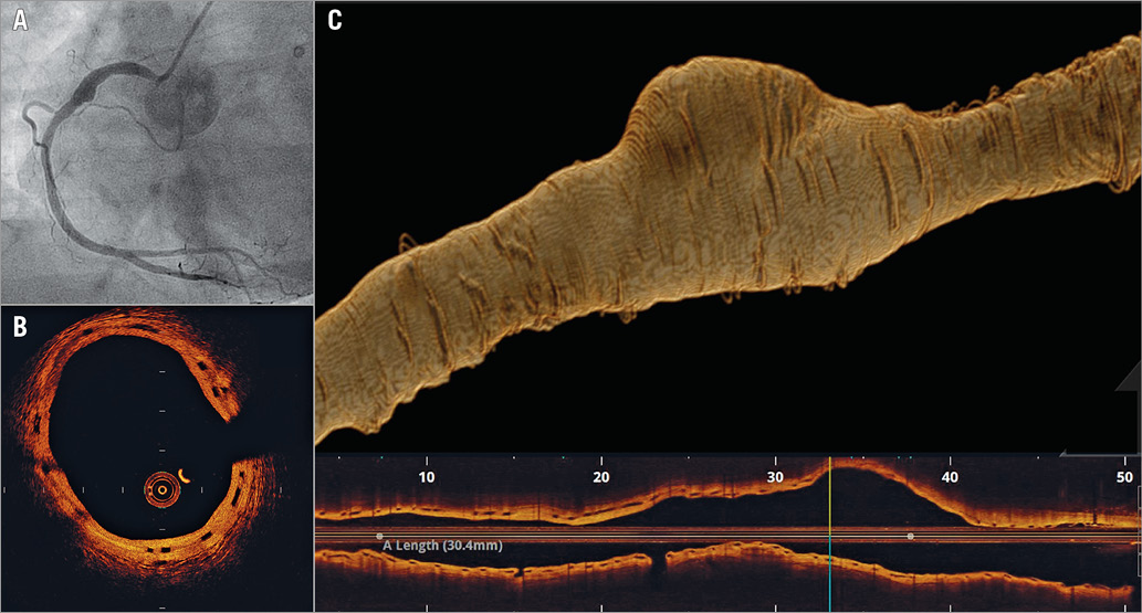
She re-presented with angina 21 months later. Angiography revealed a significant ostial RCA stenosis, with a localised coronary artery aneurysm within the scaffolded segment (Panel A). Pressure wire assessment yielded an FFR of 0.76 with a step at the ostium. Optical coherence tomography confirmed a large, acquired aneurysm at the point of minimal scaffold overlap (Panel B, Panel C). No malapposition was observed, and the scaffold appeared to have expanded with the vessel far beyond normal scaffold dimensions. A short drug-eluting stent (DES) was deployed to the ostium. The aneurysmal segment was managed conservatively with dual antiplatelet therapy (aspirin and clopidogrel).

This extreme form of positive remodelling in a BVS has not, to our knowledge, been described previously. The mechanism of acquired aneurysm following DES/BVS deployment remains unclear; however, hypersensitivity to the drug and/or polymer is possible. In this case, it is possible that “double-dose” everolimus at the point of BVS overlap may have been a factor in aneurysm formation.

Conflict of interest statement

J. Hill has received consulting fees from Abbott and St. Jude Medical. The other authors have no conflicts of interest to declare.

Volume 12 Number 9
Oct 20, 2016
Volume 12 Number 9
View full issue


Key metrics

On the same subject

IMAGE IN CARDIOLOGY

10.4244/EIJ-D-16-00323 Jan 20, 2017
Delayed fracture of a bioresorbable vascular scaffold implanted for in-stent restenosis
Bastante T et al
free

IMAGE IN CARDIOLOGY

10.4244/EIJV12I7A142 Sep 18, 2016
Plaque protrusion compromising bioresorbable coronary scaffold patency
Floré V et al
free

Image – Interventional flashlight

10.4244/EIJ-D-19-00063 Jan 20, 2021
Late structural discontinuity after bioresorbable vascular scaffold implantation in patients with in-stent restenosis
Cuesta J et al
free

Image – Interventional flashlight

10.4244/EIJ-D-18-00441 Oct 4, 2019
Long-term follow-up of an extreme kinking of a right coronary artery stenosis treated with a bioresorbable vascular scaffold
Stierlin F et al
free

IMAGE IN CARDIOLOGY

10.4244/EIJY14M10_07 Nov 20, 2015
Subacute thrombosis of a bioresorbable vascular scaffold implanted for recurrent in-stent restenosis
Rivero F et al
free
Trending articles
94.95

State-of-the-Art Review

10.4244/EIJ-D-20-01296 Aug 27, 2021
Management of cardiogenic shock
Thiele H et al
free
56.5

Clinical research

10.4244/EIJ-D-22-00621 Feb 20, 2023
Long-term changes in coronary physiology after aortic valve replacement
Sabbah M et al
free
55.1

Clinical Research

10.4244/EIJ-D-21-00875 Aug 5, 2022
Features of atherosclerosis in patients with angina and no obstructive coronary artery disease
Pellegrini D et al
free
X

The Official Journal of EuroPCR and the European Association of Percutaneous Cardiovascular Interventions (EAPCI)

EuroPCR EAPCI
PCR ESC
Impact factor: 7.6
2023 Journal Citation Reports®
Science Edition (Clarivate Analytics, 2024)
Online ISSN 1969-6213 - Print ISSN 1774-024X
© 2005-2024 Europa Group - All rights reserved