IMAGE IN CARDIOLOGY

DOI: 10.4244/EIJV12I1A10

Coronary aneurysm without malapposition after bioresorbable vascular scaffold implantation

Leo Timmers1,2*, MD, PhD; Yoke Ching Lim1, MBChB, MRCP; Huay Cheem Tan1, MBBS, MMed (Int Med), FRCP, FACC, FAMS; Adrian F. Low1, MBBS

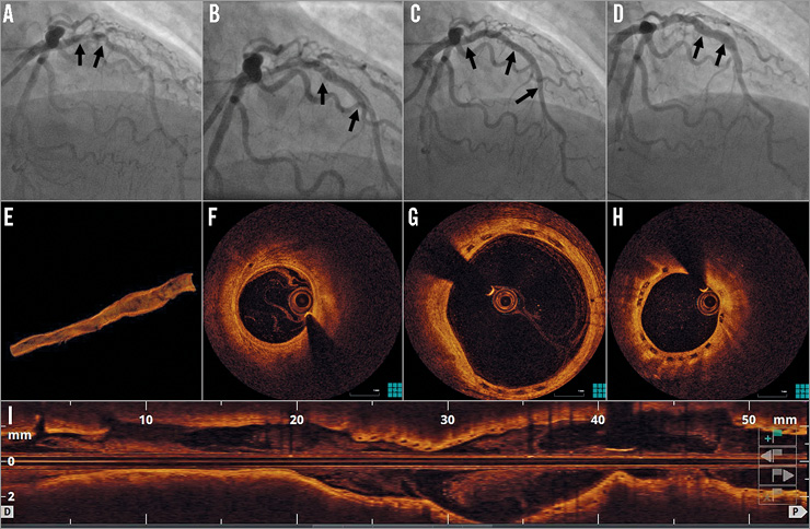
A 54-year-old male underwent coronary angiography for non-ST-elevation myocardial infarction. This identified significant left anterior descending artery (LAD) disease (Panel A, Moving image 1). After predilatation with a 3×15 mm balloon, a type B dissection occurred (Panel B, Moving image 2). Two Absorb™ bioresorbable vascular scaffolds (BVS) (Abbott Vascular, Santa Clara, CA, USA) were implanted in the mid (3×18 mm at 16 atm) and proximal (3.5×28 mm at 6 atm) LAD (Panel C, Moving image 3). After post-dilatation with a 3.5×15 mm non-compliant balloon at 18 atm, the vessel diameter was 3.5 mm. Thirty-two months later, a coronary angiogram showed a segment of ectasia in the proximal and mid LAD, with a maximal vessel diameter of 5.3 mm (Panel D, Moving image 4). Optical coherence tomography demonstrated the absence of strut malapposition. Instead, completely endothelialised strut remnants were visible in the positively remodelled vessel wall (Panel E, 3D reconstruction, Panel F, distal reference, Panel G, ectatic segment, Panel H, proximal reference, Moving image 5). This suggests that the scaffold was incorporated into the vessel wall and that gradual degradation of the scaffold resulted in disruption of strut continuity, allowing outward strut displacement along with the enlarging coronary artery. Positive remodelling causing late acquired metallic DES malapposition has been recognised as a potential cause for very late stent thrombosis. This image demonstrates that coronary aneurysm formation can occur after BVS implantation. However, in contrast to metallic DES, the transient nature of BVS integrity can result in the absence of late strut malapposition. Whether this entails a lower risk of very late thrombosis after positive coronary arterial remodelling remains uncertain.

Conflict of interest statement

The authors have no conflicts of interest to declare.

Supplementary data

Moving image 1. Baseline coronary angiogram.

Moving image 2. Coronary angiogram after balloon dilatation.

Moving image 3. Angiographic result after BVS implantation.

Moving image 4. Angiogram 32 months after BVS implantation.

Moving image 5. Optical coherence tomography.

Supplementary data

To read the full content of this article, please download the PDF.

Baseline coronary angiogram.

Coronary angiogram after balloon dilatation.

Angiographic result after BVS implantation.

Angiogram 32 months after BVS implantation.

Optical coherence tomography.

Volume 12 Number 1
May 16, 2016
Volume 12 Number 1
View full issue


Key metrics

Suggested by Cory

IMAGE IN CARDIOLOGY

10.4244/EIJV12I9A89 Oct 20, 2016
Acquired coronary artery aneurysm following treatment with bioresorbable vascular scaffolds
O’Gallagher K et al
free

IMAGE IN CARDIOLOGY

10.4244/EIJV11I1A14 May 19, 2015
Very late bioresorbable vascular scaffold thrombosis: a new clinical entity
Azzalini L et al
free

IMAGE IN CARDIOLOGY

10.4244/EIJV11I7A158 Nov 20, 2015
A very late thrombosis in a bioresorbable vascular scaffold
Giacchi M et al
free

SHORT REPORT

10.4244/EIJ-D-17-00353 Apr 6, 2018
Very late scaffold thrombosis: insights from optical coherence tomography and histopathology
Joner M et al
free

IMAGE IN CARDIOLOGY

10.4244/EIJV12I7A142 Sep 18, 2016
Plaque protrusion compromising bioresorbable coronary scaffold patency
Floré V et al
free

EXPERT REVIEW

10.4244/EIJ-D-16-00471 Feb 20, 2017
Possible mechanical causes of scaffold thrombosis: insights from case reports with intracoronary imaging
Sotomi Y et al
free

IMAGE IN CARDIOLOGY

10.4244/EIJ-D-16-00259 Mar 20, 2017
Neoatherosclerosis: an emerging and conceptually unexpected cause of very late bioresorbable vascular scaffold failure
Hiltrop N et al
free
Trending articles
200.45

State-of-the-Art

10.4244/EIJ-D-21-00089 Jun 11, 2021
Intracoronary optical coherence tomography: state of the art and future directions
Ali ZA et al
free
154.43

State-of-the-Art

10.4244/EIJ-D-22-00776 Apr 3, 2023
Computed tomographic angiography in coronary artery disease
Serruys PW et al
free
92.95

State-of-the-Art Review

10.4244/EIJ-D-20-01296 Aug 27, 2021
Management of cardiogenic shock
Thiele H et al
free
47.4

State-of-the-Art

10.4244/EIJ-D-24-00386 Feb 3, 2025
Mechanical circulatory support for complex, high-risk percutaneous coronary intervention
Ferro E et al
free
43.65

Clinical research

10.4244/EIJ-D-23-00590 Dec 4, 2023
Prognostic impact of cardiac damage staging classification in each aortic stenosis subtype undergoing TAVI
Nakase M et al
free
36.5

State-of-the-Art

10.4244/EIJ-D-23-00448 Jan 15, 2024
Coronary spasm and vasomotor dysfunction as a cause of MINOCA
Yaker ZS et al
free
34.75

State-of-the-Art

10.4244/EIJ-D-23-00606 Jan 1, 2024
Targeting inflammation in atherosclerosis: overview, strategy and directions
Waksman R et al
free
X

The Official Journal of EuroPCR and the European Association of Percutaneous Cardiovascular Interventions (EAPCI)

EuroPCR EAPCI
PCR ESC
Impact factor: 7.6
2023 Journal Citation Reports®
Science Edition (Clarivate Analytics, 2024)
Online ISSN 1969-6213 - Print ISSN 1774-024X
© 2005-2025 Europa Group - All rights reserved