IMAGE IN CARDIOLOGY

DOI: 10.4244/EIJV12I7A142

Plaque protrusion compromising bioresorbable coronary scaffold patency

Vincent Floré1*, MD, PhD; Mariano Pellicano1, MD; Julien Adjedj1, MD; Angela Ferrara1, MD; Kris Van Der Steen2, MD; Jozef Bartunek1, MD, PhD

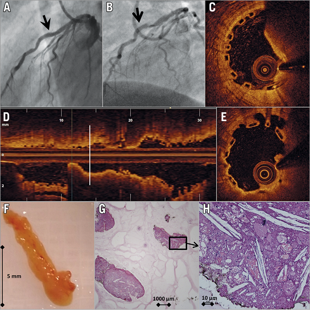
The patient was a 53-year-old (male) suffering from unstable angina due to stenosis of the left anterior descending (LAD) artery. After 2.5 mm balloon predilatation (12 atm), a 3.0×28 mm bioresorbable vascular scaffold (BVS) (Absorb™; Abbott Vascular, Santa Clara, CA, USA) was implanted at 14 atm with an angiographically good result (Panel A). The patient was discharged on aspirin and clopidogrel.

Coronary angiography one month later showed a severe lesion in the distal part of the BVS, suggesting in-stent thrombosis (Panel B). Optical coherence tomography (OCT) confirmed mixed thrombus (Panel C, Panel D). With an aspiration catheter, a white structure measuring 9×2 mm (Panel F) was evacuated. Control OCT confirmed the disappearance of a protrusive structure, revealing an area of suboptimal BVS expansion and apposition (Panel E). A 3.5 mm non-compliant balloon post-dilatation (16 atm) was performed. OCT re-evaluation after one month was inconspicuous.

Platelet reactivity test (Multiplate® analyser; Roche Diagnostics Limited, Rotkreuz, Switzerland) demonstrated normal response to clopidogrel. Histologic examination of the aspirated debris revealed atherosclerotic plaque with a high concentration of cholesterol crystals, histiocytes, lymphocytes and red blood cells (Panel G, Panel H).

Although suggestive of in-scaffold thrombosis, this image shows the probable cause to be plaque protrusion into the scaffold lumen. The mechanism is eccentric plaque position in combination with insufficient expansion and apposition of a BVS within an angulated segment. This case emphasises the need for lesion preparation and post-implant optimisation when implanting BVS.

Conflict of interest statement

The authors have no conflicts of interest to declare.

Volume 12 Number 7
Sep 18, 2016
Volume 12 Number 7
View full issue


Key metrics

Suggested by Cory

IMAGE IN CARDIOLOGY

10.4244/EIJV11I1A14 May 19, 2015
Very late bioresorbable vascular scaffold thrombosis: a new clinical entity
Azzalini L et al
free

EXPERT REVIEW

10.4244/EIJ-D-16-00471 Feb 20, 2017
Possible mechanical causes of scaffold thrombosis: insights from case reports with intracoronary imaging
Sotomi Y et al
free

IMAGE IN CARDIOLOGY

10.4244/EIJY14M10_07 Nov 20, 2015
Subacute thrombosis of a bioresorbable vascular scaffold implanted for recurrent in-stent restenosis
Rivero F et al
free

SHORT REPORT

10.4244/EIJ-D-17-00353 Apr 6, 2018
Very late scaffold thrombosis: insights from optical coherence tomography and histopathology
Joner M et al
free

IMAGE IN CARDIOLOGY

10.4244/EIJV11I7A158 Nov 20, 2015
A very late thrombosis in a bioresorbable vascular scaffold
Giacchi M et al
free

IMAGE IN CARDIOLOGY

10.4244/EIJV10I5A103 Sep 28, 2014
Acute Absorb bioresorbable vascular scaffold thrombosis in ST-segment elevation myocardial infarction: to stent or not to stent?
Fernández-Rodríguez D et al
free

IMAGE IN CARDIOLOGY

10.4244/EIJ-D-16-00259 Mar 20, 2017
Neoatherosclerosis: an emerging and conceptually unexpected cause of very late bioresorbable vascular scaffold failure
Hiltrop N et al
free
Trending articles
87.2

State-of-the-Art

10.4244/EIJ-D-25-00266 Jan 19, 2026
Lesion stratification with intracoronary imaging
McGarvey M et al
free
47.45

NEW INNOVATION

10.4244/EIJ-D-15-00467 Feb 20, 2018
Design and principle of operation of the HeartMate PHP (percutaneous heart pump)
Van Mieghem NM et al
free
34.8

Original Research

10.4244/EIJ-D-25-01006 Mar 16, 2026
Clinical outcomes and haemodynamic response after blinded stress assessment of moderate aortic stenosis
Eerdekens R et al
22.2

Viewpoint

10.4244/EIJ-D-25-01066 May 4, 2026
Intracoronary imaging guidance for de novo coronary lesion treatment with drug-coated balloons
Amabile N et al
free
20.75

Flashlight

10.4244/EIJ-D-25-01014 Apr 6, 2026
Stent retriever-assisted coronary thrombectomy with continuous aspiration
Liabot Q et al
open access
19.5

Original Research

10.4244/EIJ-D-26-00032 May 15, 2026
Glucocorticoids to reduce permanent pacemaker implantation after TAVI: the GLUCO-TAVI randomised trial
Fuertes-Kenneally L et al
17.8

Expert Review

10.4244/EIJ-D-25-01316 Apr 20, 2026
Electrosurgical laceration and stabilisation of tricuspid edge-to-edge repair: the ELASTA-T technique
Alvarez-Covarrubias H et al
free
X

PCR
Impact factor: 9.5
2024 Journal Citation Reports®
Science Edition (Clarivate Analytics, 2025)
Online ISSN 1969-6213 - Print ISSN 1774-024X
© 2005-2026 Europa Group - All rights reserved