IMAGE IN CARDIOLOGY

DOI: 10.4244/EIJY14M10_07

Subacute thrombosis of a bioresorbable vascular scaffold implanted for recurrent in-stent restenosis

Fernando Rivero, MD; Amparo Benedicto, MD; Teresa Bastante, MD; Javier Cuesta, MD; Guillermo Diego, MD; Fernando Alfonso*, MD

An 88-year-old diabetic man was admitted for progressive angina. Four years before, he required a bare metal stent (BMS) implantation in a severely calcified lesion at the ostium of the right coronary artery. Six months later he presented severe focal in-stent restenosis (ISR) which was treated with a new BMS. Urgent coronary angiography showed recurrent ISR (Figure 1A). Optical coherence tomography (OCT) revealed the classic “stent-sandwich” image with two well-expanded stents (Figure 1B). The tissue causing ISR was highly heterogeneous, suggestive of neoatherosclerosis (Figure 1B). After pre-treatment with 600 mg of clopidogrel, aggressive lesion dilation was performed. Then, a bioresorbable vascular scaffold (BVS) (3.5×12 mm) was implanted and post-dilated with a 3.75 mm non-compliant balloon at 26 bar. OCT revealed good results but with some persistent areas of BVS underexpansion (Figure 1C).

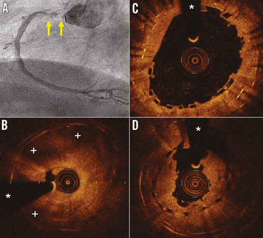
Figure 1. Angiographic and intravascular findings. A) Angiography showing recurrent severe in-stent restenosis of the proximal right coronary artery (arrows). B) Optical coherence tomography disclosing obstructive neoatherosclerosis with large lipidic areas (+). C) Optical coherence tomography image obtained immediately after BVS post-dilation. The black box appearance of the device is visualised over a double metal stent layer. D) Optical coherence tomography image at the time of BVS thrombosis. The obstructive thrombus is readily recognised but also a clear recoil of the BVS was detected (intra-scaffold minimal area: distal 5.3 mm2 [Δ=–1.5 mm2]; proximal 4 mm2 [Δ=–1.1 mm2]). (*) denotes wire artefact.

Thirty-six hours later, the patient experienced chest pain and the ECG revealed inferior ST-segment elevation. Angiography showed occlusion at the right coronary ostium. OCT disclosed a mixed thrombus obstructing the BVS (Figure 1D). Surprisingly, the BVS minimal lumen area was significantly reduced compared with results immediately after the procedure, demonstrating the occurrence of subacute BVS recoil (Moving image 1). Aggressive multiple balloon dilatations were performed and eventually a satisfactory final result was obtained.

Conflict of interest statement

The authors have no conflicts of interest to declare.

Supplementary data

Moving image 1. BVS subacute thrombosis.

Volume 11 Number 7
Nov 20, 2015
Volume 11 Number 7
View full issue


Key metrics

On the same subject

IMAGE IN CARDIOLOGY

10.4244/EIJV12I7A142 Sep 18, 2016
Plaque protrusion compromising bioresorbable coronary scaffold patency
Floré V et al
free

10.4244/EIJV11I12A279 Mar 18, 2016
Recurrent in-stent restenosis: many treatment options, no certainty
Piraino D et al
free

Image – Interventional flashlight

10.4244/EIJ-D-19-00063 Jan 20, 2021
Late structural discontinuity after bioresorbable vascular scaffold implantation in patients with in-stent restenosis
Cuesta J et al
free

Image – Interventional flashlight

10.4244/EIJ-D-20-00507 Aug 6, 2021
Optical coherence tomography and tailored treatment of in-stent restenosis
Jia H et al
free

SHORT REPORT

10.4244/EIJ-D-17-00353 Apr 6, 2018
Very late scaffold thrombosis: insights from optical coherence tomography and histopathology
Joner M et al
free

10.4244/EIJV11I12A259 Mar 18, 2016
Oversized post-dilatation of current bioresorbable vascular scaffolds: kill or cure?
von Birgelen C and Basalus M
free
Trending articles
338.63

State-of-the-Art Review

10.4244/EIJ-D-21-00904 Apr 1, 2022
Antiplatelet therapy after percutaneous coronary intervention
Angiolillo D et al
free
295.45

Expert consensus

10.4244/EIJ-D-21-00898 Sep 20, 2022
Intravascular ultrasound guidance for lower extremity arterial and venous interventions
Secemsky E et al
free
226.03

State-of-the-Art Review

10.4244/EIJ-D-21-00426 Dec 3, 2021
Myocardial infarction with non-obstructive coronary artery disease
Lindahl B et al
free
209.5

State-of-the-Art Review

10.4244/EIJ-D-21-01034 Jun 3, 2022
Management of in-stent restenosis
Alfonso F et al
free
168.4

Expert review

10.4244/EIJ-D-21-00690 May 15, 2022
Crush techniques for percutaneous coronary intervention of bifurcation lesions
Moroni F et al
free
149.53

State-of-the-Art

10.4244/EIJ-D-22-00776 Apr 3, 2023
Computed tomographic angiography in coronary artery disease
Serruys PW et al
free
103.48

Expert consensus

10.4244/EIJ-E-22-00018 Dec 4, 2023
Definitions and Standardized Endpoints for Treatment of Coronary Bifurcations
Lunardi M et al
free
X

The Official Journal of EuroPCR and the European Association of Percutaneous Cardiovascular Interventions (EAPCI)

EuroPCR EAPCI
PCR ESC
Impact factor: 6.2
2022 Journal Citation Reports®
Science Edition (Clarivate Analytics, 2023)
Online ISSN 1969-6213 - Print ISSN 1774-024X
© 2005-2024 Europa Group - All rights reserved