IMAGE IN CARDIOLOGY

DOI: 10.4244/EIJV10I12A252

Two-month healing evaluation of an everolimus Pt-Cr DES with erodible polymer and two bioresorbable scaffolds implanted in the same vessel of the same patient

Jose M. de la Torre Hernandez*, MD, PhD, FESC Dae-Hyun Lee, MD Tamara Garcia Camarero, MD Fermin Sainz Laso, MD Ines Toranzo, MD Leire Goicolea, MD Javier Zueco, MD

A 43-year-old male underwent primary angioplasty over the left circumflex artery and was scheduled to have lesions in the left anterior descending artery (LAD) treated days later (Appendix Figure 1A). The intention was to treat all lesions with bioresorbable scaffolds but the most distal lesion had to be covered with a lower profile 2.25/38 mm everolimus Pt-Cr stent (SYNERGY™; Boston Scientific, Marlborough, MA, USA). Two 2.5/28 mm Absorb bioresorbable vascular scaffolds (BVS) (Abbott Vascular, Santa Clara, CA, USA) were implanted in the mid and proximal segments (Appendix Figure 1B).

The patient was admitted two months later for unstable angina showing progression of a previously moderate lesion in the right coronary artery which was treated. We evaluated the LAD with optical coherence tomography.

In the SYNERGY stent, a complete and smooth coverage was observed over all struts (Figure 1A). In the BVS, the degree of coverage was high but not complete (Figure 1B). The difference between the two could be related mostly to the large difference in strut thickness (74 µm vs. 150 µm). Nevertheless, we could not distinguish the neointimal or fibrin components of the thin strut-covering layers.

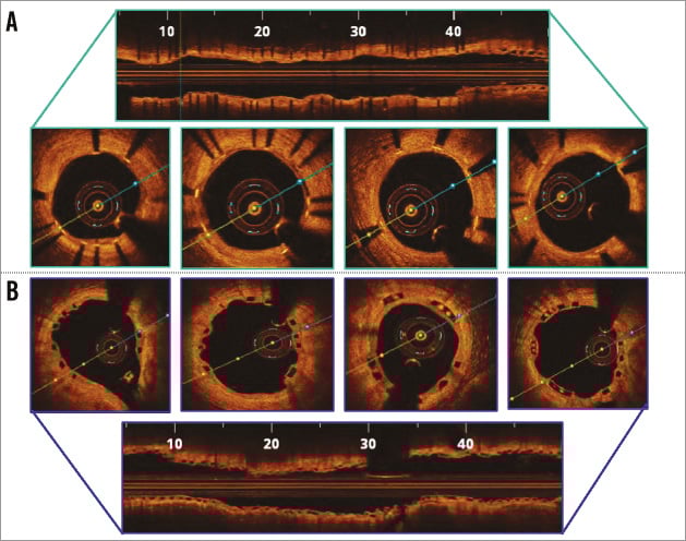
Figure 1. OCT findings two months after implantation. A) Everolimus Pt-Cr (SYNERGY) stent. B) Bioresorbable scaffolds (BVS).

Conflict of interest statement

J. M. de la Torre Hernandez receives research grants from Abbott Vascular, Boston Scientific, Biotronik, Biosensors, and St. Jude. He also receives payments for presentations and/or advisory board membership from Abbott, Boston Scientific, Biotronik, Medtronic, Biosensors, Volcano, Lilly, AstraZeneca. The other authors have no conflicts of interest to declare.

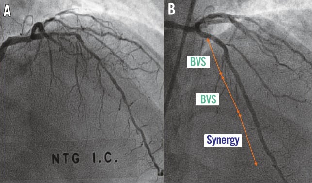
Appendix Figure 1. Angiographic images before and after stent implantation. A) Angiography showing lesions in the LAD. B) Angiographic result after stent implantation.

Volume 10 Number 12
Apr 20, 2015
Volume 10 Number 12
View full issue


Suggested by Cory

IMAGE IN CARDIOLOGY

10.4244/EIJV12I7A142 Sep 18, 2016
Plaque protrusion compromising bioresorbable coronary scaffold patency
Floré V et al
free

IMAGE IN CARDIOLOGY

10.4244/EIJV11I1A14 May 19, 2015
Very late bioresorbable vascular scaffold thrombosis: a new clinical entity
Azzalini L et al
free
Trending articles
58.3

State-of-the-Art

10.4244/EIJ-D-24-00386 Feb 3, 2025
Mechanical circulatory support for complex, high-risk percutaneous coronary intervention
Ferro E et al
free
39.45

Clinical research

10.4244/EIJ-D-23-00725 Nov 19, 2023
A systematic algorithm for large-bore arterial access closure after TAVI: the TAVI-MultiCLOSE study
Rosseel L et al
free
39.45

Original Research

10.4244/EIJ-D-23-00725 Mar 18, 2024
A systematic algorithm for large-bore arterial access closure after TAVI: the TAVI-MultiCLOSE study
Rosseel L et al
free
36.25

State-of-the-Art

10.4244/EIJ-D-23-00448 Jan 15, 2024
Coronary spasm and vasomotor dysfunction as a cause of MINOCA
Yaker ZS et al
free
35.15

State-of-the-Art

10.4244/EIJ-D-23-00895 Apr 1, 2024
Percutaneous interventions for pulmonary embolism
Finocchiaro S et al
free
28.5

CLINICAL RESEARCH

10.4244/EIJV11I1A6 May 19, 2015
European expert consensus on rotational atherectomy
Barbato E et al
free
X

The Official Journal of EuroPCR and the European Association of Percutaneous Cardiovascular Interventions (EAPCI)

EuroPCR EAPCI
PCR ESC
Impact factor: 7.6
2023 Journal Citation Reports®
Science Edition (Clarivate Analytics, 2024)
Online ISSN 1969-6213 - Print ISSN 1774-024X
© 2005-2025 Europa Group - All rights reserved