IMAGE IN CARDIOLOGY

DOI: 10.4244/EIJ-D-16-00323

Delayed fracture of a bioresorbable vascular scaffold implanted for in-stent restenosis

Teresa Bastante, MD; Javier Cuesta, MD; Fernando Rivero, MD; Marcos García-Guimaraes, MD; Amparo Benedicto, MD; Fernando Alfonso*, MD, PhD

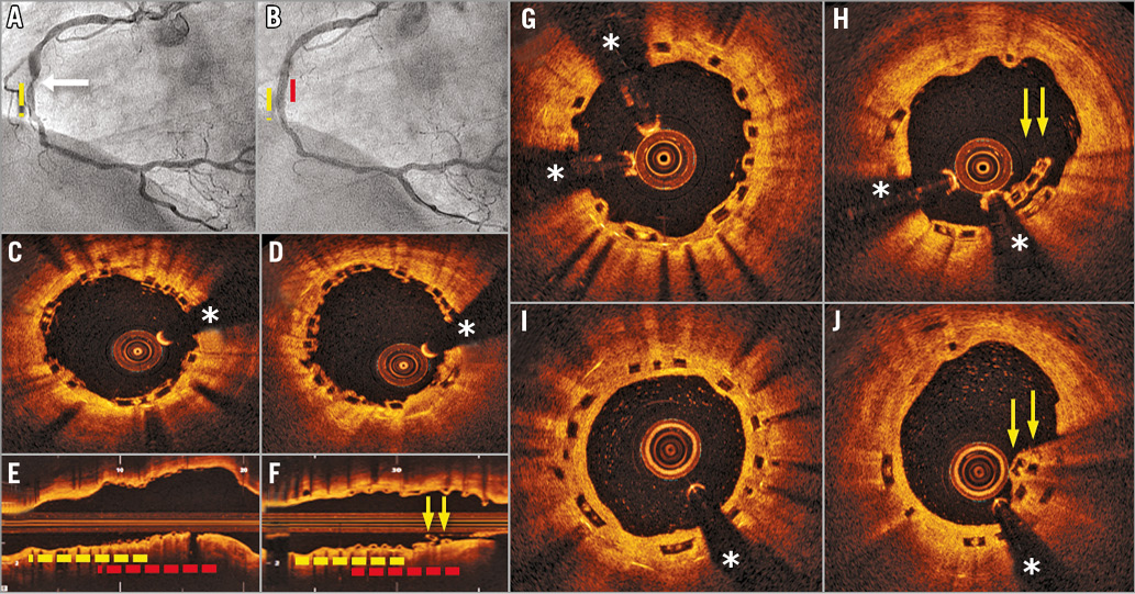
A 61-year-old woman presented with effort angina one year after the implantation of a drug-eluting stent (DES) in the mid right coronary artery (RCA). Coronary angiography revealed a proximal “edge” in-stent restenosis (Panel A, arrow; yellow broken line: DES) that was successfully treated with a 3×8 mm bioresorbable vascular scaffold (BVS) (Absorb™; Abbott Vascular, Santa Clara, CA, USA) at 20 bar (Panel B, red broken line: BVS). Optical coherence tomography (OCT) confirmed an excellent expansion and apposition of the BVS (Panel C-Panel E, yellow broken line: DES; red broken line: BVS). One month later she was admitted for an inferior acute myocardial infarction. Urgent coronary angiography showed a good result of the BVS but complete occlusion of the most distal RCA that was treated with a new DES. On OCT, the BVS was covered by incipient bright neointima. However, a localised malapposition of some BVS struts was readily identified leading to the diagnosis of late scaffold fracture (Panel F-Panel H, arrows). No further intervention was made. At eight-month follow-up, the BVS maintained an excellent angiographic result and OCT revealed complete neointimal coverage of the fractured BVS (Panel I, Panel J, arrows) (*denotes wire artefact).

Stent fracture is a rare complication described in metallic stents and, more recently, also in BVS. Stent fracture is associated with major complications including restenosis, aneurysm/pseudoaneurysm formation and thrombosis. Risk factors include high implantation pressures and hinge point location. Our BVS was implanted at high pressures but not at a hinge point. The fracture was detected well before the time required for resorption-induced structural BVS disintegrity. Iatrogenic mechanical disruption can never be completely discarded but no clues were identified in this regard. Notably, the BVS rupture was located precisely at the edge of the previous DES. The potential interaction of the rigid underlying DES platform with the flexible overlapping BVS structure should be investigated. To the best of our knowledge, this is the first description of a delayed fracture in a BVS implanted for in-stent restenosis.

Conflict of interest statement

The authors have no conflicts of interest to declare.

Volume 12 Number 13
Jan 20, 2017
Volume 12 Number 13
View full issue


Key metrics

Suggested by Cory

Image – Interventional flashlight

10.4244/EIJ-D-19-00063 Jan 20, 2021
Late structural discontinuity after bioresorbable vascular scaffold implantation in patients with in-stent restenosis
Cuesta J et al
free

IMAGE IN CARDIOLOGY

10.4244/EIJV11I1A14 May 19, 2015
Very late bioresorbable vascular scaffold thrombosis: a new clinical entity
Azzalini L et al
free

IMAGE IN CARDIOLOGY

10.4244/EIJY14M10_07 Nov 20, 2015
Subacute thrombosis of a bioresorbable vascular scaffold implanted for recurrent in-stent restenosis
Rivero F et al
free

IMAGE IN CARDIOLOGY

10.4244/EIJV12I1A10 May 16, 2016
Coronary aneurysm without malapposition after bioresorbable vascular scaffold implantation
Timmers L et al
free

EXPERT REVIEW

10.4244/EIJ-D-16-00471 Feb 20, 2017
Possible mechanical causes of scaffold thrombosis: insights from case reports with intracoronary imaging
Sotomi Y et al
free
Trending articles
309.93

State-of-the-Art Review

10.4244/EIJ-D-21-00695 Nov 19, 2021
Transcatheter treatment for tricuspid valve disease
Praz F et al
free
166.7

Expert review

10.4244/EIJ-D-21-00690 May 15, 2022
Crush techniques for percutaneous coronary intervention of bifurcation lesions
Moroni F et al
free
92.2

State-of-the-Art Review

10.4244/EIJ-D-20-01296 Aug 27, 2021
Management of cardiogenic shock
Thiele H et al
free
76

State-of-the-Art

10.4244/EIJ-D-23-00840 Sep 2, 2024
Aortic regurgitation: from mechanisms to management
Baumbach A et al
free
72.85

State-of-the-Art

10.4244/EIJ-D-24-00386 Feb 3, 2025
Mechanical circulatory support for complex, high-risk percutaneous coronary intervention
Ferro E et al
free
58.3

Clinical research

10.4244/EIJ-D-23-00344 Sep 18, 2023
Clinical outcomes of TAVI with the Myval balloon-expandable valve for non-calcified aortic regurgitation
Sanchez-Luna JP et al
free
56.65

Clinical research

10.4244/EIJ-D-20-01155 Oct 20, 2021
A deep learning algorithm for detecting acute myocardial infarction
Liu W et al
free
33.9

CLINICAL RESEARCH

10.4244/EIJ-D-17-00381 Oct 11, 2017
Stent malapposition and the risk of stent thrombosis: mechanistic insights from an in vitro model
Foin N et al
free
33.65

State-of-the-Art

10.4244/EIJ-D-23-00606 Jan 1, 2024
Targeting inflammation in atherosclerosis: overview, strategy and directions
Waksman R et al
free
X

The Official Journal of EuroPCR and the European Association of Percutaneous Cardiovascular Interventions (EAPCI)

EuroPCR EAPCI
PCR ESC
Impact factor: 7.6
2023 Journal Citation Reports®
Science Edition (Clarivate Analytics, 2024)
Online ISSN 1969-6213 - Print ISSN 1774-024X
© 2005-2025 Europa Group - All rights reserved