IMAGE IN CARDIOLOGY

DOI: 10.4244/EIJ-D-16-00384

Usefulness of three-dimensional optical frequency domain imaging for diagnosing in-stent restenosis due to a stent fracture

Yusuke Oba, MD; Nobuhiko Ogata*, MD, PhD; Motoki Fukutomi, MD, PhD; Kazuomi Kario, MD, PhD

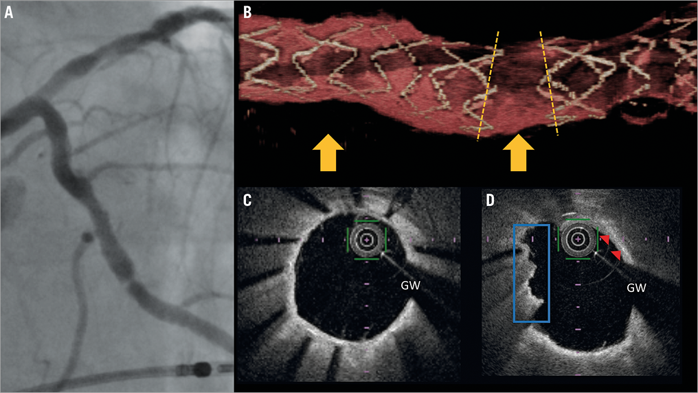
A 74-year-old man underwent a percutaneous coronary intervention (PCI) due to worsening effort angina pectoris. We implanted a 3.0×18 mm DES (Nobori®; Terumo Corp., Tokyo, Japan) in the left circumflex coronary artery (LCx). The final angiographic results were satisfactory for the targeted lesion. Thirty-three months later, coronary angiography revealed in-stent restenosis (Panel A, Moving image 1). After an angioplasty using a 3.0×15 mm balloon (Lacrosse® NSE ALPHA™; Goodman Co., Ltd, Nagoya, Japan), optical frequency domain imaging (OFDI) was performed. The multiple struts of the stent were completely fractured at the acquired transection with a gap in the stent body (Panel B, Moving image 2). Although Panel C shows circumferentially covered stent struts, superficial high-intensity high-attenuation plaque and a hazy appearance by OFDI might suggest accumulations of macrophage and mural thrombus on this fractured site (Panel D). Therefore, the main reason for in-stent restenosis in this case might be persistent mechanical stress due to total separation type stent fracture. Finally, an angioplasty was performed with a 3.0×15 mm paclitaxel-coated balloon (SeQuent® Please; B. Braun, Melsungen, Germany).

Acknowledgement

We acknowledge the kind advice received from Dr Gaku Nakazawa.

Conflict of interest statement

The authors have no conflicts of interest to declare.

Supplementary data

Moving image 1. Follow-up angiography revealed in-stent restenosis in the LCx.

Moving image 2. 3-dimensional OFDI showed the total separation type stent fracture.

Supplementary data

To read the full content of this article, please download the PDF.

video1
video2
Volume 12 Number 11
Dec 9, 2016
Volume 12 Number 11
View full issue


Key metrics

On the same subject

IMAGE IN CARDIOLOGY

10.4244/EIJ-D-16-00323 Jan 20, 2017
Delayed fracture of a bioresorbable vascular scaffold implanted for in-stent restenosis
Bastante T et al
free

Image – Interventional flashlight

10.4244/EIJ-D-19-00063 Jan 20, 2021
Late structural discontinuity after bioresorbable vascular scaffold implantation in patients with in-stent restenosis
Cuesta J et al
free

IMAGE IN CARDIOLOGY

10.4244/EIJV12I10A201 Nov 20, 2016
Ruptured “non-culprit” in-stent neoatherosclerosis during ST-segment elevation acute myocardial infarction
Cuesta J et al
free

Image – Interventional flashlight

10.4244/EIJ-D-20-00507 Aug 6, 2021
Optical coherence tomography and tailored treatment of in-stent restenosis
Jia H et al
free

State-of-the-Art Review

10.4244/EIJ-D-21-01034 Jun 3, 2022
Management of in-stent restenosis
Alfonso F et al
free

Clinical research

10.4244/EIJ-D-23-00107 Aug 7, 2023
Mechanisms and treatment outcomes of ostial right coronary artery in-stent restenosis
Yamamoto K et al

IMAGE IN CARDIOLOGY

10.4244/EIJV12I6A121 Aug 20, 2016
Optical coherence tomography visualisation of burst balloon catheter trapped by coronary stent
Shimura T et al
free

Image – Interventional flashlight

10.4244/EIJ-D-21-00393 Apr 22, 2022
Napkin ring formation in culotte stenting using current-generation drug-eluting stents in left main coronary artery bifurcation
Murasato Y et al
free
Trending articles
281.53

State-of-the-Art Review

10.4244/EIJ-D-21-00695 Nov 19, 2021
Transcatheter treatment for tricuspid valve disease
Praz F et al
free
243.2

State of the art

10.4244/EIJ-D-21-01117 Sep 20, 2022
Recanalisation of coronary chronic total occlusions
Di Mario C et al
free
208.35

State-of-the-Art Review

10.4244/EIJ-D-21-01034 Jun 3, 2022
Management of in-stent restenosis
Alfonso F et al
free
168.7

Translational research

10.4244/EIJ-D-21-00824 May 15, 2022
Bench test and in vivo evaluation of longitudinal stent deformation during proximal optimisation
Toth GG et al
free
167.05

Expert review

10.4244/EIJ-D-21-00690 May 15, 2022
Crush techniques for percutaneous coronary intervention of bifurcation lesions
Moroni F et al
free
151.03

State-of-the-Art

10.4244/EIJ-D-22-00776 Apr 3, 2023
Computed tomographic angiography in coronary artery disease
Serruys PW et al
free
118

Translational research

10.4244/EIJ-D-22-00718 Jun 5, 2023
Preclinical evaluation of the degradation kinetics of third-generation resorbable magnesium scaffolds
Seguchi M et al
110.35

Viewpoint

10.4244/EIJ-E-22-00007 May 15, 2022
TAVI at 20: how a crazy idea led to a clinical revolution
Eltchaninoff H et al
free
X

The Official Journal of EuroPCR and the European Association of Percutaneous Cardiovascular Interventions (EAPCI)

EuroPCR EAPCI
PCR ESC
Impact factor: 6.2
2022 Journal Citation Reports®
Science Edition (Clarivate Analytics, 2023)
Online ISSN 1969-6213 - Print ISSN 1774-024X
© 2005-2024 Europa Group - All rights reserved