A 77-year-old man was admitted to our institution with a lateral ST-segment elevation myocardial infarction. Two years earlier, a paclitaxel-eluting stent (PES) and an everolimus-eluting stent (EES) had been implanted separately in the left circumflex coronary artery. Clopidogrel was discontinued after 12 months; aspirin was maintained indefinitely. Coronary angiography proved definite stent thrombosis (ST). After manual thrombus aspiration restenosis involving both stents was observed (Figure 1A). Optical coherence tomography (OCT) detected in-stent heterogeneous, low-intensity and attenuated neointima (Figure 1B, Figure 1C, Figure 1F, Figure 1G, Moving image 1, Moving image 2) compared with the homogeneous, high-intensity signal of adjacent fibrous tissue (Figure 1D, Figure 1E). After balloon angioplasty, the in-stent “sponge-like” pattern changed to a thin-layer neointima, similar to the signal from the interposed vessel segment (Appendix Figure 1, Appendix Figure 2).
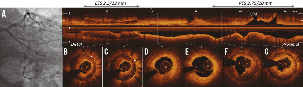
Figure 1. Angiography and OCT after manual thrombus aspiration. Severe in-stent restenosis was observed in the PES and EES (A, dotted lines). OCT demonstrated heterogeneous, signal-poor and attenuated neointima in both stents (B, C and F, G) compared to a homogeneous and intense signal in-between (D, E). Rich neovascularisation was detected within neointima (B, C, arrowheads).
Atherosclerotic changes of the neointima may act as a causative factor for late ST. The present case demonstrates that neoatherosclerosis (NA) can occur also in newer-generation drug-eluting stents. The high lipid content and the soft tissue characterising NA make a balloon angioplasty a possible therapeutic option to avoid multilayered stent implantation.
Conflict of interest statement
G. Guagliumi and V. Sirbu received consulting/grant support from St. Jude Medical. The other authors have no conflicts of interest to declare.
Supplementary data
Moving image 1. OCT pullback pre balloon angioplasty focused on EES. OCT pullback shows characteristics of the EES neoatherosclerosis.
Moving image 2. OCT pullback pre balloon angioplasty focused on PES. OCT pullback shows characteristics of the PES neoatherosclerosis.
Supplementary data
To read the full content of this article, please download the PDF.
OCT pullback pre balloon angioplasty focused on EES. OCT pullback shows characteristics of the EES neoatherosclerosis.
Moving image 2. OCT pullback pre balloon angioplasty focused on PES. OCT pullback shows characteristics of the PES neoatherosclerosis.