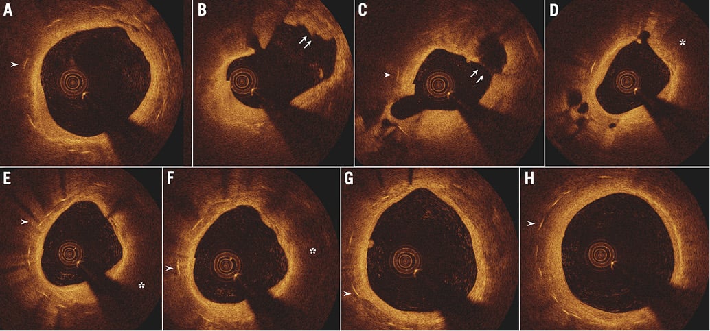
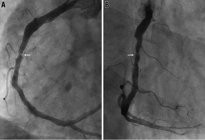
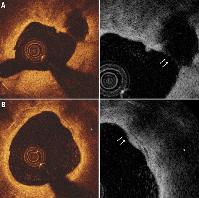
In-stent neoatherosclerosis has recently been described as an important mechanism of late stent failure. A 56-year-old man with a single bare metal stent implantation ten years previously presented with new-onset chest pain. Coronary angiography revealed a right coronary artery with focal in-stent restenosis (Online Figure 1). Optical coherence tomography (OCT) demonstrated the presence of fibroatheroma throughout the entire length of the stent. At one point (panel C), a thin-cap fibroatheroma (TCFA) with plaque rupture is noted (Figure 1, Online Figure 2).

Figure 1. The OCT images from distal (A) to proximal (H). Arrowheads indicate stent struts. A lipid-rich (asterisk) plaque with a TCFA has developed inside the stent. At the site of plaque rupture (C), there is a large empty cavity leaving a residual fibrous cap behind (arrows). Notice an intraluminal thrombus (B, arrows).
Conflict of interest statement
The authors have no conflicts of interest to declare.

Online Figure 1. Right coronary artery in left (A) and right (B) anterior oblique.

Online Figure 2. Magnified OCT images. Disrupted intima with residual fibrous cap with empty cavity (A, arrows). Intact fibroatheroma with fibrous cap thickness of 60 μm (B). The images on the right are visualised as inverse greyscales.

