IMAGE IN CARDIOLOGY

DOI: 10.4244/EIJV10I12A251

Very late stent thrombosis caused by neoatherosclerotic plaque rupture

Mikkel Hougaard*, MD; Per Thayssen, MD, DMSci; Henrik Steen Hansen, MD, DMSci; Lisette Okkels Jensen, MD, PhD, DMSci

A 51-year-old man with non-ST-segment elevation myocardial infarction was admitted for coronary angiography which revealed thrombotic occlusion in a segment containing two overlapping CYPHER® stents (Cordis, Johnson & Johnson, Bridgewater, NJ, USA) implanted two years earlier. The patient had a family history of ischaemic heart disease and was previously treated with percutaneous coronary intervention in the right coronary artery and in the circumflex artery (Cx). Optical coherence tomography was performed prior to predilatation and showed well-apposed stents, complete coverage and in-stent hyperplasia with neoatherosclerosis, especially in the distal part (Figure 1A). A plaque rupture containing white thrombus material was identified (Figure 1B) and more proximally we found deposits of white and red thrombus material. Stent struts covering the origin of the first marginal branch were covered and free of thrombus material. A third stent more proximal in the Cx showed evagination but full coverage without evidence of thrombus.

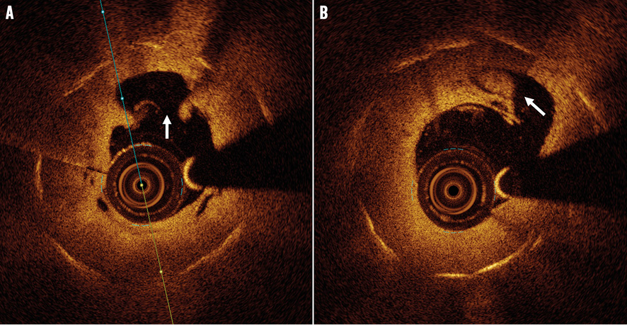
Figure 1. OCT findings. A) Intimal hyperplasia with plaque rupture (arrow). B) White thrombus in relation to rupture site (arrow).

The case demonstrates rupture in a neoatherosclerotic plaque as a mechanism behind very late stent thrombosis (Appendix Figures).

Conflict of interest statement

L.O. Jensen has received an unrestricted research grant to the institution from St. Jude Medical. The other authors have no conflicts of interest to declare.

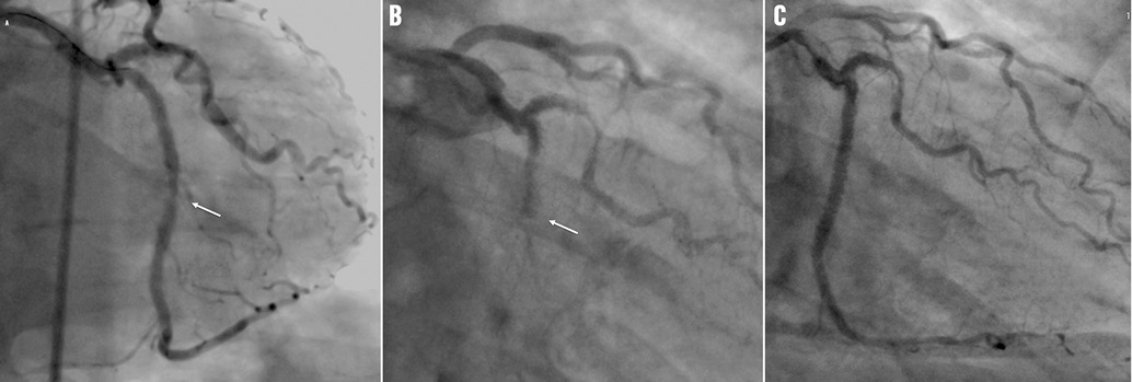
Appendix Figure 1. Coronary angiogram findings. A) Narrowing of the Cx seen in relation to earlier PCI of RM1 (arrow). B) Thrombotic occlusion of the Cx (arrow). C) Final result after PCI.

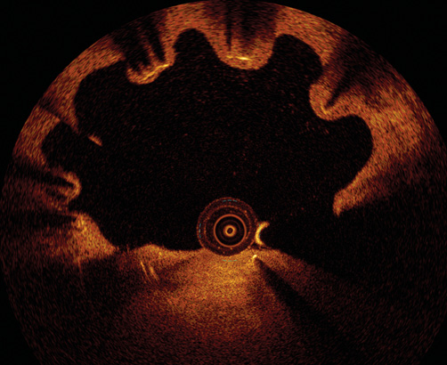
Appendix Figure 2. OCT findings. Malapposed but covered stent struts in the more proximal stent in the Cx possibly due to evagination.

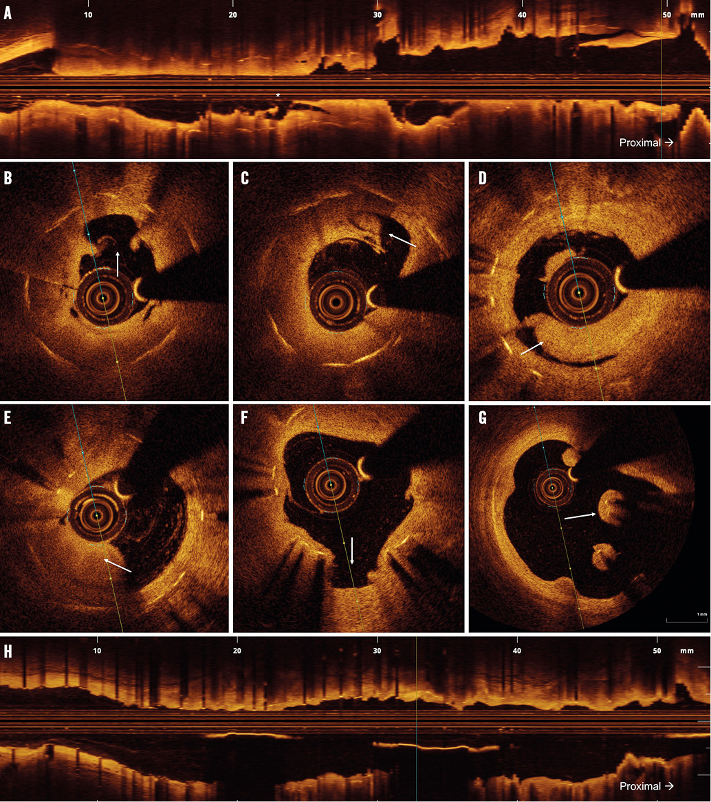
Appendix Figure 3. OCT findings. A) Longitudinal overview of the thrombosed segment. Blood in the proximal part (*) marks the plaque rupture site. B) Intimal hyperplasia with plaque rupture (arrow). C) & D) White thrombus in relation to the rupture site (arrow). E) Red thrombus proximal to the rupture site (arrow). F) Thrombus occluding minor side branch. G) Malapposed but covered stent struts at the origin of RM1. H) Longitudinal overview after additional stenting.

Online data supplement

Moving image 1. OCT pullback prior to PCI.

Moving image 2. OCT pullback after PCI.

Supplementary data

To read the full content of this article, please download the PDF.

Moving image 1.
Moving image 2.
Volume 10 Number 12
Apr 20, 2015
Volume 10 Number 12
View full issue


Key metrics

Suggested by Cory

IMAGE IN CARDIOLOGY

10.4244/EIJV12I10A201 Nov 20, 2016
Ruptured “non-culprit” in-stent neoatherosclerosis during ST-segment elevation acute myocardial infarction
Cuesta J et al
free

IMAGE IN CARDIOLOGY

10.4244/EIJV11I7A157 Nov 20, 2015
Stent failure due to simultaneous aggressive neoatherosclerosis of first- and current-generation drug-eluting stents
Komukai K et al
free

Image – Interventional flashlight

10.4244/EIJ-D-20-00507 Aug 6, 2021
Optical coherence tomography and tailored treatment of in-stent restenosis
Jia H et al
free

Image – Interventional flashlight

10.4244/EIJ-D-20-00731 Aug 6, 2021
Periprocedural stent thrombosis after percutaneous coronary intervention for a bifurcation lipid-rich plaque
Häner JD et al
free
Trending articles
318.1

State-of-the-Art Review

10.4244/EIJ-D-21-00695 Nov 19, 2021
Transcatheter treatment for tricuspid valve disease
Praz F et al
free
116.75

State-of-the-Art

10.4244/EIJ-D-24-00066 Apr 21, 2025
Management of complications after valvular interventions
Bansal A et al
free
108.3

Viewpoint

10.4244/EIJ-E-22-00007 May 15, 2022
TAVI at 20: how a crazy idea led to a clinical revolution
Eltchaninoff H et al
free
91.6

Image – Interventional flashlight

10.4244/EIJ-D-22-00344 Aug 5, 2022
First dedicated transcatheter leaflet splitting device: the ShortCut device
Tchétché D et al
free
72.4

State-of-the-art

10.4244/EIJ-D-22-00627 Feb 6, 2023
Left atrial appendage occlusion
Holmes D et al
free
68.9

State-of-the-Art

10.4244/EIJ-D-24-00992 Sep 15, 2025
Antithrombotic therapy in complex percutaneous coronary intervention
Castiello D et al
free
49.55

CLINICAL RESEARCH

10.4244/EIJ-D-17-00962 Apr 6, 2018
A new optical coherence tomography-based calcium scoring system to predict stent underexpansion
Fujino A et al
free
43.45

State-of-the-Art Review

10.4244/EIJ-D-21-00145 Sep 20, 2021
Robotics, imaging, and artificial intelligence in the catheterisation laboratory
Beyar R et al
free
X

PCR
Impact factor: 9.5
2024 Journal Citation Reports®
Science Edition (Clarivate Analytics, 2025)
Online ISSN 1969-6213 - Print ISSN 1774-024X
© 2005-2025 Europa Group - All rights reserved