A 59-year-old man with ST-segment elevation myocardial infarction was admitted for primary percutaneous coronary intervention due to a severe stenosis in the proximal left anterior descending artery (LAD) at the bifurcation of the first diagonal branch (Medina type 1,1,1) with TIMI flow 2 in both branches (Panel A). After thrombus aspiration and pre-dilatation of the side branch and the main vessel, the LAD was stented with a 3.0×23mm sirolimus-eluting stent (Cypher Select+; Cordis, Miami Lakes, FL, USA), resulting in severe ostial side branch (SB) stenosis (Panel B). To facilitate SB access, proximal optimisation technique (POT)1 was performed by dilating the proximal segment with a short 3.5×8mm non-compliant balloon. After rewiring the SB, wire position was controlled by optical coherence tomography (OCT) (C7; St. Jude Medical, St. Paul, MN, USA) and 3D-OCT images were reconstructed by use of an OCT-XA registration software package, prototype version = (Medis medical imaging systems bv, Leiden, The Netherlands)2. The side branch wire was found to pass through the stent lumen into the narrow SB ostium without passing under proximal stent struts (Panel C). Final kissing balloon dilatation (FKBD) was performed by simultaneously inflating 3.0×15mm non-compliant balloons at 14 atm (Panel D). 3D-OCT after FKBD showed adequate opening of the SB ostium and some distortion (ratio of longest/shortest stent diameter=1.4) of the proximal part of the stent was seen (Panel E). The longest diameter in the oval shaped proximal main vessel stent was in the same plane as the SB offspring due to FKBD with parallel balloons. The ostia of both distal vessels were visible from the proximal end of the stent (PanelE). This could indicate a favourable anatomy facilitating flow into both distal branches.
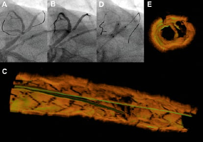
Figure 1. A) Coronary angiogram pre PCI; B) After main vessel stenting; C) 3D-OCT showing wirer position in jailed side branch; D) Final kissing balloon dilatation with parallel balloons; 3D-OCT showing proximal stent distortion and opening of ostia after kissing balloon dilatation. See text for details.
This is the first time highly informative 3D-OCT images have been reconstructed in a few steps by a novel OCT-XA software package. In this case the 3D-OCT images provided evidence of correct re-wiring and successful FKBD. A larger series is warranted to provide further insight into optimal re-wiring of the SB and subsequent outcome after FKBD.
Conflict of interest statement
N.Holm received speaker fees, travel grants and unrestricted research grants from St. Jude Medical and a research grant from Cordis. S. Tu has a research appointment at the Leiden University Medical Center and is employed by Medis medical imaging systems bv. E. Christiansen has received speaker fees and a travel grant from Cordis. J. Reiber has a part-time appointment as professor of medical imaging at the Leiden University Medical Center and is CEO of Medis medical imaging systems bv. J.Lassen has recieved speaker fees and unrestricted research grants from Cordis and st. Jude Medical. L. Thuesen has recieved unrestricted reseach grants form Cordis. M. Maeng has received travel grants and speaker fees from Cordis.