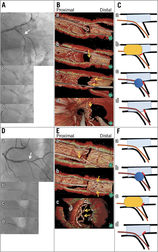

For the optimal treatment of the side branch (SB) after main vessel (MV) stenting in coronary bifurcation lesions, confirming the guidewire recrossing (GWR) point in the SB ostium is crucial1. Three-dimensional (3D) imaging with optical coherence tomography (OCT) can visualise the GWR point clearly. We encountered unusual deflection of the protruded struts treated with the proximal optimisation technique (POT) and SB dilation, when the GWR into proximal cells of the struts overhung into the SB ostium.
Deflection to the SB
A 67-year-old man with a 1,1,0 lesion in the left circumflex artery obtuse marginal branch bifurcation (Panel Aa), in which vessel references in the proximal, distal MV and SB were 3.2, 2.6, and 2.8 mm, respectively, underwent zotarolimus-eluting 2.75×12 mm stent (Medtronic, Minneapolis, MN, USA) implantation at 8 atm (Panel Ab). POT was performed with the stent delivery balloon at 12 atm with its distal marker located in the carina to ensure that the stent was well apposed (Panel Ac). A 2.5×4 mm Glider PTCA balloon (TriReme, Pleasanton, CA, USA) was subsequently dilated in the SB ostium (Panel Ad). The procedure was guided with two-dimensional (2D) OCT (St. Jude Medical, St. Paul, MN, USA) and the data were sent to another hospital for 3D reconstruction using dedicated software (INTAGE Realia; CYBERNET, Tokyo, Japan). The 3D image demonstrated GWR into the proximal cell (Panel Ba); however, the protruded struts were folded towards the distal SB after SB dilation (Panel Bb-Bd, Moving image 1, Moving image 2).
Deflection to the distal MV
An 80-year-old man with a 1,1,0 lesion in the distal right coronary artery (Panel Da), in which vessel references in the proximal, distal main vessels and side branch were 2.8, 2.3, and 2.5 mm, respectively, underwent zotarolimus-eluting 2.25×26 mm stent implantation (Panel Db). GWR and subsequent SB dilation with a 2.5×4 mm Glider balloon were performed (Panel Dc), followed by POT dilation with a 3.25×10 mm balloon (Panel Dd). The 3D OCT image also demonstrated GWR into the proximal cell (Panel Ea); however, the protruded struts turned towards the distal MV after SB dilation (Panel Eb, Panel Ec). The deflected struts were apposed to the vessel with final kissing balloon inflation (Moving image 3).
These two bifurcation interventions were performed similarly, except for the sequence of POT and SB dilation; however, deflection of the protruded struts at the carinal area was in opposite directions. In case 1, POT before SB dilation enabled adequate strut expansion with some deflection towards the distal SB, and subsequent SB dilation completed this deflection (Panel C). In contrast, in case 2, POT after SB dilation resulted in deflection of the protruded struts towards the distal MV (Panel F). The remaining more obstructive struts after SB dilation following the zotarolimus-eluting stent deployment2 might enhance the risk of deflection of the protruded struts. POT should be performed before SB dilation, and 3D OCT is helpful to identify the GWR point and avoid serious stent deformation.
Conflict of interest statement
J. Shite is a compensated consultant for St. Jude Medical. The other authors have no conflicts of interest to declare.
Supplementary data
Moving image 1. Flythrough view from proximal to distal MV in case 1.
Moving image 2. Flythrough view from proximal MV to SB in case 1.
Moving image 3. Flythrough view from proximal to distal MV in case 2.
To read the full content of this article, please download the PDF.
Supplementary data
To read the full content of this article, please download the PDF.
Moving image 1. Flythrough view from proximal to distal MV in case 1.
Moving image 2. Flythrough view from proximal MV to SB in case 1.
Moving image 3. Flythrough view from proximal to distal MV in case 2.

