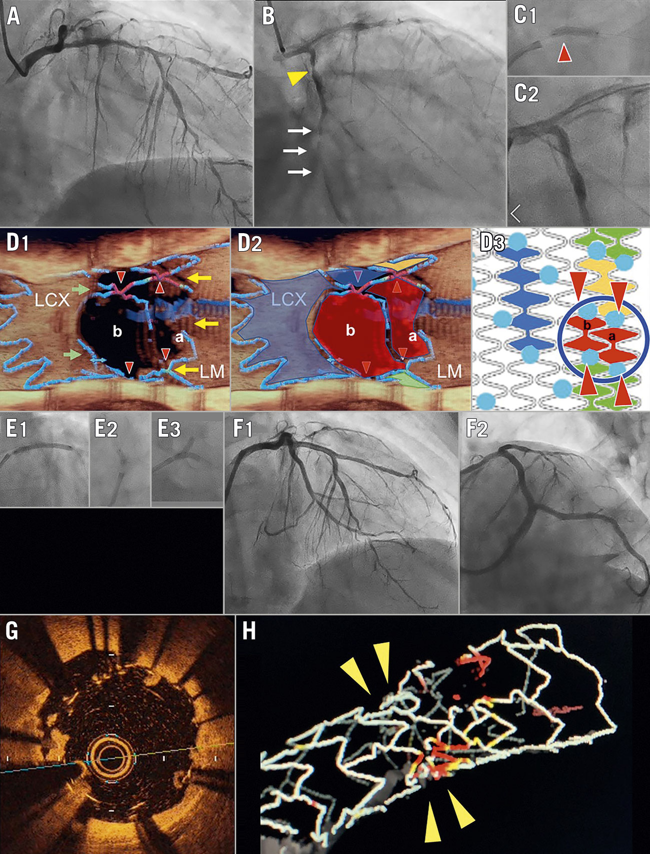
Figure 1. Case presentation. A) Baseline coronary angiography (CAG): RAO cranial view. B) RAO caudal view showing a subtotal occlusion (arrows) and 90% stenosis in the LCX (yellow triangle). C) Second PCI, 1 week later. C1) Resolute Onyx (3.0/15 mm) implantation in the LCX with protrusion into the LM (red triangle). C2) CAG failed to show the stent-jailed LAD. D) Third PCI, 1 month later. D1) 3D-OCT image at the LAD ostium: jailing struts with four link connections (red triangles), and guidewire crossing the most proximal cell (a). D2) Stent edge with the smallest cells (red) occupied. D3) Design of Resolute Onyx stent. The stent edge aligns smaller cells with fewer crowns (crown no.: red, 2; orange, 3; green, 4; blue [regular], 5) and more link connections (blue dots). The circle indicates the location of LAD ostium. E) LM stenting. E1) Culotte stenting with a 3.0 mm stent deployment. E2) LAD and LM dilation with a 4.0 mm balloon. E3) KBI with 3.0 and 2.5 mm balloons. F) Final CAG. F1) RAO cranial view. F2) LAO caudal view. G) Malapposed struts with thrombus attachment. H) Napkin ring formation of the stents. CAG: coronary angiography; KBI: kissing balloon inflation; LAD: left anterior descending artery; LCX: left circumflex artery; LM: left main; PCI: percutaneous coronary intervention; RAO: right anterior oblique; 3D-OCT: three-dimensional optical coherence tomography
A 64-year-old man with non-ST-elevation myocardial infarction underwent primary stenting in the middle left circumflex artery (LCX) (Figure 1A, Figure 1B, Moving image 1, Moving image 2). One week later, the diseased right coronary artery was treated with stent implantation. Then the operator attempted to implant a 3.0/15 mm Resolute Onyx stent (Medtronic) at the proximal LCX, but failed due to the patient’s deep breathing and accidental disengagement of the guiding catheter, causing it to protrude into the left main (LM) (Figure 1C1). Proximal optimisation and kissing balloon inflation (KBI) in the LM bifurcation (Figure 1C2) resulted in complete attachment of the proximal stent edge to the LM and some opening of the jailed left anterior descending artery (LAD) (Moving image 3, Moving image 4). In the intervention on the LAD one month later, an optical coherence tomography (OCT) catheter (Dragonfly; Abbott) could not be advanced towards the LAD. The three-dimensional OCT (3D-OCT) image of pullback from the LCX revealed that the protruded struts and four link connections (red triangles) of the LCX stent had jailed the LAD ostium with the guidewire crossing into the most proximal cell (Figure 1D1, Figure 1D2, cell a). Small spaces for wiring remained outside the stent for stent crush (yellow arrows) and inside the distal regular cell for cell expansion (green arrows). After successful rewiring to the distal cell (cell b), a 3.0/12 mm non-compliant balloon was inflated at 22 atm. As the medium size of the Resolute Onyx stent has more link connections and smaller cells with 2-4 crowns in the proximal site (fewer than the 5 crowns in regular cells), in order to maintain helical coil constitution (Figure 1D3), two consecutive smaller cells (red cell a, red cell b) with maximal expansion capacities <3.0 mm were aligned at the LAD ostium (Figure 1D2). We implanted a XIENCE Alpine™ 3.0/38 mm stent (Abbott Laboratories) from the LM to the LAD to form minimal culotte stenting (Figure 1E1). The LM and the proximal LAD were dilated with a 4.0/8 mm non-compliant balloon (Figure 1E2) and subsequent KBI with 3.0 mm and 2.5 mm balloons at 12 atm was performed (Figure 1E3). The final angiography showed acceptable results (Figure 1F, Moving image 5, Moving image 6). However, OCT showed a limited stent expansion area of 5.3 mm2 and remarkable malapposition, with a range of 400-700 μm, combined with thrombus attachment at the LAD ostium (Figure 1G, Moving image 7). Napkin ring formation was demonstrated on 3D-OCT images (Figure 1H, Moving image 8).
Although distal cell wiring and subsequent jailed branch dilation is the standard technique1, maximal cell expansion is dependent on the stent platform (number of crowns and links), rewiring site, and link position at the ostium23. Current-generation drug-eluting stents with fewer link connections and greater cell expansion capacity rarely present a napkin ring formation in culotte stenting2. However, alignment of smaller cells at the stent edge is applied by the Resolute Onyx, Orsiro (Biotronik), and SYNERGY (Boston Scientific) stents. When these smaller cells jail the branch ostium, napkin ring formation is likely to occur, as in the present case. As the proximal stent edge reached as far as the LM, the other option for guidewire crossing outside of the LCX stent (Figure 1D1, yellow arrows) and performing double kissing crush was technically difficult. Cells a and b folding back in the LCX ostium would lead to similar expansion restriction, even after KBI2. Other preventive or bail-out procedures are summarised in Supplementary Appendix 1. Although minimising site overlap is recommended in two-stenting techniques1, caution is needed to avoid jailing these stents by smaller cells.
Conflict of interest statement
The authors have no conflicts of interest to declare.
Supplementary data
To read the full content of this article, please download the PDF.
Moving image 1. Baseline coronary angiography (right anterior oblique [RAO] cranial view).
Moving image 2. Baseline coronary angiography (RAO caudal view).
Moving image 3. Final intravascular ultrasound (IVUS) image in the second percutaneous coronary intervention (PCI): pullback from the LCX to the LM.
Moving image 4. Final IVUS image in the second PCI: pullback from the LAD to the LM.
Moving image 5. Final coronary angiography in the third PCI (RAO cranial view).
Moving image 6. Final coronary angiography in the third PCI (LAO caudal view).
Moving image 7. OCT pullback from the LAD.
Moving image 8. Three-dimensional OCT image of the LAD stent with a napkin ring formation at the LAD artery ostium.