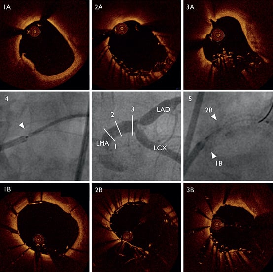
Coronary angiography of a 71-year-old male patient with unstable angina revealed subtotal distal left main artery (LMA) stenosis extending into the proximal left anterior descending artery (LAD) and 80% obstruction of the origin of the proximal left circumflex artery (LCX). The patient was treated with T-stenting at the bifurcation of the left anterior descending and left circumflex. During the procedure profound longitudinal stent compression was noticed, followed by OCT. (Figure 1) This novel complication is of great concern for operators and has the potential to affect current treatment of ostial lesions.

Figure 1. T-stenting of the bifurcation was scheduled starting with a PROMUS Element™ stent (PES; Boston Scientific, Natick, MA, USA) (4.0/28 mm, 24 atm) at the ostium of the LMA (4) reaching into the proximal LAD, extended into the proximal and mid LAD with a second PES. During deployment of this distal stent, the ostial segment of the proximal stent was compressed longitudinally (2A, multiple strut layers) leaving a dissection at the ostium (1A), and more than two thirds of the LMA uncovered. Instead of the intended T-stenting a Resolute Integrity™ stent (RIS; Medtronic, Minneapolis, MN, USA) (4.0/15 mm) was placed in culotte technique, starting at the ostium (1B, 5) to cover the dissection and stent-free proximal LMA, reaching into the LCX. Optical coherence tomography was performed immediately after longitudinal compression (top row) and after placement of the RIS (bottom row). Accumulation of multiple strut layers reaching far into the lumen (3A). Double stent layer, improved apposition of compressed PES segment (2B, 5). Reduced strut accumulation (3B).
Conflict of interest statement
G. Leibundgut has received a fellowship grant from Biotronik and consulting fees from Abbott Laboratories. M. Gick has received lecture fees from Boston Scientific. None of the other authors have any conflicts of interest to declare.
Online data supplement
Moving image 1. Optical coherence tomography (OCT) Recording of proximal left anterior descending artery (LAD) and left main artery (LMA).
Supplementary data
To read the full content of this article, please download the PDF.
Moving image 1. Optical coherence tomography (OCT) Recording of proximal left anterior descending artery (LAD) and left main artery (LMA).

