A 40-year-old male patient wounded by a bomb fragment during the war in the Middle East was admitted to our hospital complaining of a persistent cough. On a three-dimensional CT scan, a pseudoaneurysm on the proximal part of the left common carotid artery and a fistula between its distal level and brachiocephalic vein were detected. Furthermore, the anomalous origin of the left common carotid artery from the brachiocephalic trunk was noteworthy (Figure 1). The diagnosis was confirmed with carotid angiography (Moving image 1, Moving image 2). In another session, a 6 Fr guiding catheter with 0.035 inch J guidewire from the right femoral artery was used to enter the left common carotid artery. At the same time, the right radial approach with a 5 Fr pigtail catheter was used for angiography and to determine the ostium of the left common carotid artery (Moving image 3). A 10/50 mm GORE® VIABAHN® graft stent (Gore Medical, Flagstaff, AZ, USA) was implanted to 2 mm distal from the ostium of the left common carotid artery, covering the pseudoaneurysm and the fistula area (Moving image 4, Moving image 5). On control CT angiography, the treatment was deemed successful (Figure 1).
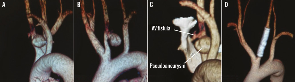
Figure 1. 3D CT scan of carotid artery before and after intervention. A) & B) The three-dimensional CT scan revealed a pseudoaneurysm on the proximal part of the left common carotid artery. In addition, the anomalous origin of the left common carotid from the brachiocephalic trunk was noteworthy. C) Three-dimensional CT scan with a pseudoaneurysm on the proximal part of the left common carotid artery and a fistula between its distal level and brachiocephalic vein. D) Control CT angiography after treatment.
Conflict of interest statement
The authors have no conflicts of interest to declare.
Online data supplement
Moving image 1. Carotid angiography.
Moving image 2. Carotid angiography.
Moving image 3. Carotid angiography.
Moving image 4. Intervention on carotid artery.
Moving image 5. Intervention on carotid artery.
Supplementary data
To read the full content of this article, please download the PDF.
Carotid angiography.
Moving image 2. Carotid angiography.
Moving image 3. Carotid angiography.
Moving image 4. Intervention on carotid artery.
Moving image 5. Intervention on carotid artery.