IMAGE IN CARDIOLOGY

DOI: 10.4244/EIJV12I7A143

Unexpected entrapment of a guidewire by bioresorbable vascular scaffold deployment at a calcified coronary lesion

Wei-Chieh Lee, MD; Hsiu-Yu Fang, MD; Pei-Hsun Sung, MD; Kuo-Ho Yeh, MD; Chiung-Jen Wu*, MD

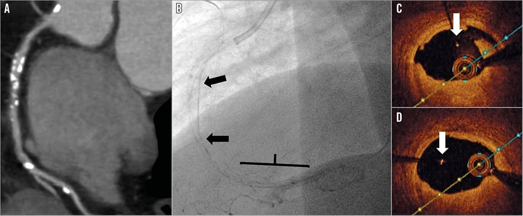
Entrapment of a broken guidewire after percutaneous coronary intervention is rare, with a reported incidence of 0.2%. This case involved a 64-year-old man with a high coronary calcified score (Agatston/Janowitz score: 817) by multislice cardiac computed tomography (Panel A). Intravascular ultrasound showed surrounding calcified plaque at the mid right coronary artery (RCA). Predilation was carried out and a long dissection was noted at the mid RCA with flow limitation. However, a bioresorbable vascular scaffold (BVS) (Absorb™; Abbott Vascular, Santa Clara, CA, USA) could not pass smoothly through the tight calcified lesion. A buddy wire technique was used with a 0.014” Runthrough® NS Floppy (Terumo Corp., Tokyo, Japan) and a 0.014” Fielder FC (ASAHI Intecc, Aichi, Japan) wire. One 3.0×28 mm BVS was deployed at the distal RCA followed by in-stent high-pressure dilatation using a 3.0×15 mm Hiryu® non-compliant PTCA balloon catheter (Terumo) at 20 atm. The guidewire got stuck in the distal scaffold edge while being pulled back. A Finecross® microcatheter (Terumo) and small balloon were used to create a channel to try to pull out the guidewire but in vain. However, the distal portion of the guidewire was broken and left over the proximal to mid RCA (Panel B [arrows], Moving image 1). A 2.5×23 mm DES (XIENCE V®; Abbott Vascular) was deployed at the proximal posterior lateral branch due to the dissection. Another BVS (3.5×28 mm) was deployed at the proximal RCA, and a DES (XIENCE V, 3.5×23 mm) was deployed at the ostial RCA. Optical coherence tomography showed a well-expanded BVS at the proximal RCA, and the floating guidewire at the mid RCA (Panel C [arrow], Panel D [arrow], Moving image 2). To the best of our knowledge, this is a first case report about entrapment of a guidewire occurring after BVS deployment in a calcified lesion.

Conflict of interest statement

The authors have no conflicts of interest to declare.

Supplementary data

Moving image 1. Coronary angiography. Broken guidewire left over the proximal to mid RCA and a dissection lesion noted at the proximal RCA.

Moving image 2. Optical coherence tomography. Well-expanded BVS, floating guidewire, and multiple calcified plaque.

Supplementary data

To read the full content of this article, please download the PDF.

Coronary angiography. Broken guidewire left over the proximal to mid RCA and a dissection lesion noted at the proximal RCA.

Optical coherence tomography. Well-expanded BVS, floating guidewire, and multiple calcified plaque.

Volume 12 Number 7
Sep 18, 2016
Volume 12 Number 7
View full issue


Key metrics

Suggested by Cory

IMAGE IN CARDIOLOGY

10.4244/EIJV12I6A121 Aug 20, 2016
Optical coherence tomography visualisation of burst balloon catheter trapped by coronary stent
Shimura T et al
free

CLINICAL RESEARCH

10.4244/EIJY14M06_02 Jul 20, 2015
Extended use of the GuideLiner in complex coronary interventions
Chan P et al
free

10.4244/EIJV11SVA42 May 19, 2015
Treatment of bioresorbable scaffold failure
Felix C et al
free

IMAGE IN CARDIOLOGY

10.4244/EIJ-D-16-00323 Jan 20, 2017
Delayed fracture of a bioresorbable vascular scaffold implanted for in-stent restenosis
Bastante T et al
free

Image – Interventional flashlight

10.4244/EIJ-D-19-00063 Jan 20, 2021
Late structural discontinuity after bioresorbable vascular scaffold implantation in patients with in-stent restenosis
Cuesta J et al
free

10.4244/EIJV10I4A91 Aug 19, 2014
How should I treat an undeployed stent stuck in the proximal LAD? Going for another round...
Locca D and Eeckhout E
free

10.4244/EIJV8I7A131 Nov 20, 2012
Radial stent deformation in saphenous vein graft
Sachdeva R et al
free

10.4244/EIJV6I2A44 Jun 30, 2010
The GuideLiner™ "child" catheter
Rao U et al
free
Trending articles
87.2

State-of-the-Art

10.4244/EIJ-D-25-00266 Jan 19, 2026
Lesion stratification with intracoronary imaging
McGarvey M et al
free
47.45

NEW INNOVATION

10.4244/EIJ-D-15-00467 Feb 20, 2018
Design and principle of operation of the HeartMate PHP (percutaneous heart pump)
Van Mieghem NM et al
free
34.8

Original Research

10.4244/EIJ-D-25-01006 Mar 16, 2026
Clinical outcomes and haemodynamic response after blinded stress assessment of moderate aortic stenosis
Eerdekens R et al
22.2

Viewpoint

10.4244/EIJ-D-25-01066 May 4, 2026
Intracoronary imaging guidance for de novo coronary lesion treatment with drug-coated balloons
Amabile N et al
free
20.75

Flashlight

10.4244/EIJ-D-25-01014 Apr 6, 2026
Stent retriever-assisted coronary thrombectomy with continuous aspiration
Liabot Q et al
open access
19.5

Original Research

10.4244/EIJ-D-26-00032 May 15, 2026
Glucocorticoids to reduce permanent pacemaker implantation after TAVI: the GLUCO-TAVI randomised trial
Fuertes-Kenneally L et al
17.8

Expert Review

10.4244/EIJ-D-25-01316 Apr 20, 2026
Electrosurgical laceration and stabilisation of tricuspid edge-to-edge repair: the ELASTA-T technique
Alvarez-Covarrubias H et al
free
X

PCR
Impact factor: 9.5
2024 Journal Citation Reports®
Science Edition (Clarivate Analytics, 2025)
Online ISSN 1969-6213 - Print ISSN 1774-024X
© 2005-2026 Europa Group - All rights reserved