The mechanical function of a stent deployed in a stenosis is to provide a metallic tubular mesh scaffold to restore blood flow to perfuse the tissues downstream. We report a case of radial stent with an Export® AP Aspiration Catheter (Medtronic, Minneapolis, MN, USA) aspiration in a saphenous vein graft (SVG).
A 64-year-old man underwent coronary angiography for post-infarction angina. Angiography showed total occlusion of left anterior descending (LAD), circumflex and right coronary arteries. SVG to obtuse marginal and internal mammary graft to LAD were patent. SVG to diagonal had 90% stenosis with TIMI-1 flow. SVG to right posterior descending artery (RPDA) had 90% stenosis in the body. The patient underwent coronary intervention to SVG to diagonal with resolution of symptoms. During the same hospitalisation, his symptoms recurred requiring IV nitroglycerine and angiography was repeated. The recently placed stent was patent. A MULTI-LINKVISION™ 2.75×23 mm stent (Abbott Vascular, Abbott Park, IL, USA) was deployed at 20 atmospheres in SVG to RPDA using the GuardWire® distal protection system (Medtronic, Minneapolis, MN, USA). The stent was completely expanded on angiogram (Figure 1A and Figure 1B). During the first antegrade pass with the aspiration Export catheter, the stent was noticed to be completely expanded (Moving image1). During the Export catheter pullback, the distal 3-4 mm of the stent collapsed towards the catheter resulting in stent deformation (Figure 1C and Moving image 2). Optical coherence tomography (OCT) was performed which showed a D-shaped to oval-shaped underexpanded stent at the distal end (Figure 1D, Figure 1E, Figure 1F). Post 3.0×12 mm balloon catheter dilatation repeat OCT showed better stent apposition.
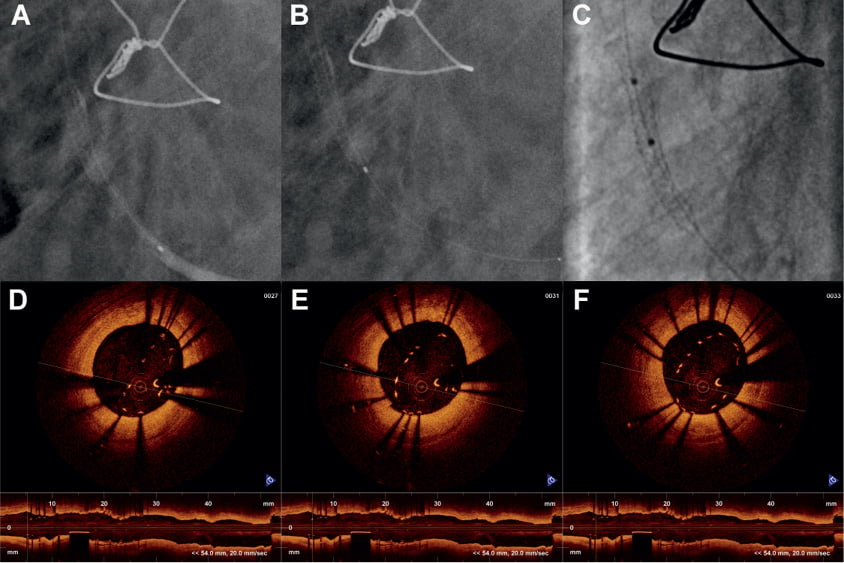
Figure 1. A) Angiographic still frame showing completely expanded stent post deployment in saphenous vein graft and first pass of the Export catheter aspiration. The GuardWire balloon is occlusive and there is a column of contrast distal to the stent with Export catheter radio-opaque marker above the contrast column. B) Angiographic still frame showing radiopaque marker of the Export catheter near the distal end of the stent as the catheter is pulled out. Note that the stent is still completely expanded. C) Angiographic still frame showing the collapse of the distal 3-4 mm of the stent post aspiration with Export catheter. Two markers of the 3.0 balloon catheter in the proximal segment of the stent are seen. D, E and F) Three optical coherence tomography frames taken from the collapsed terminal portion of the stent in both longitudinal and radial views (frame on the left with the maximal collapse is the distal terminal of the stent). In panel D, the maximal collapse is from the 6 o’clock to the 1 o’clock position giving the expanded stent a “D” shape and in the other two frames it is an oval shape. The struts are more or less symmetrically apart and not clustered or missing on one side.
Our case is unique because the distal end of the stent facing the coronary bed collapsed during Export catheter pullback with the blunt bevelled end of the catheter facing downwards. The deformed stent struts were symmetrically apart suggesting vein graft collapse with negative suction as the possible mechanism rather than the Export catheter getting caught in the stent struts.
Conflict of interest statement
R. Sachdeva serves on the Speakers Bureau for Volcano Corporation and St. Jude Medical. The other authors have no conflicts of interest to declare.
Online data supplement
Moving image 1. This shows the completely deployed stent during the antegrade passing of the Export catheter for aspiration.
Moving image 2. This shows the collapsing of the terminal end of the stent while retracting the Export catheter with continued negative suctioning.