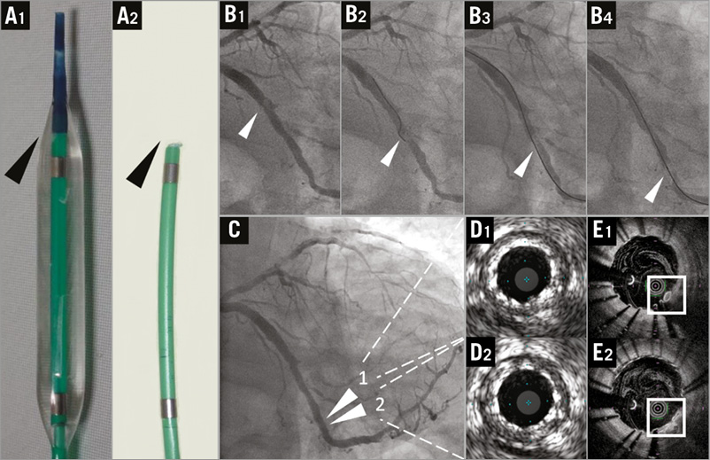

A 69-year-old man with prior anterior myocardial infarction underwent percutaneous coronary intervention for significant stenosis of the left anterior descending (LAD) artery. High-pressure inflation by non-compliant balloon was required because of severe calcification in the culprit lesion. The balloon ruptured during inflation and an angiographic filling defect appeared in the LAD. The balloon catheter was pulled out of the body and the tip and membranous part of the catheter was lost (Panel A, black arrowhead). Although we attempted to retrieve the foreign body in the LAD using a gooseneck snare, grasping the small fragment was impossible. Then we carefully pulled a guidewire whilst clasping the balloon tip. On the way, the tip unfortunately dropped and moved into the left circumflex (LCx) artery. Instead of removing it, we tried to push it into the moderate stenotic lesion at the distal LCx using a stiff guidewire (Panel B) and press it against the vessel wall with a coronary stent. After the stent implantation, angiograms showed disappearance of lumen stenosis and filling defect in the LCx (Panel C). Although the burst balloon tip could not be detected with 20 MHz intravascular ultrasound (IVUS) images, optical coherence tomography (OCT) could visualise the balloon tip between the vessel wall and stent struts (Panel D, Panel E, white squares, Moving image 1, Moving image 2). High-resolution OCT images confirmed successful bail-out from impracticable removal of the small foreign body (Online Figure 1). The clinical course of the patient was favourable and there have been no adverse events such as stent thrombosis.
Conflict of interest statement
The authors have no conflicts of interest to declare.
Supplementary data

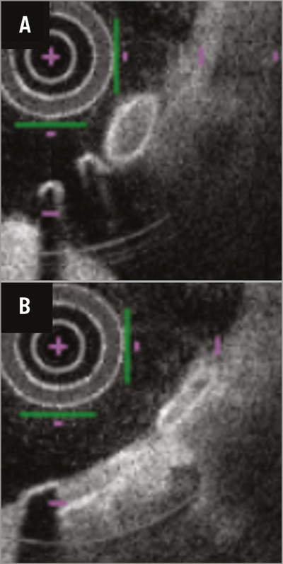
Online Figure 1. Magnified OCT images of the foreign body. Magnified OCT images of the white squares in Panel E of the image in the main article. Long diameters of ovals (0.46 mm in panel A and 0.54 mm in panel B) approximate to the diameter of the balloon tip (0.44 mm). Although there is narrow space between the stent struts and the vessel wall in panel A, complete apposition of the struts is found in panel B. Moreover, the difference in the oval shape suggests ideal stent compression in the cross-section of panel B. The transparent membrane of the balloon is not clearly visualised by OCT.
Moving image 1. OCT reveals a small foreign body between the vessel wall and the stent struts.
Moving image 2. A magnified OCT image shows compression of the foreign body by the struts. The findings suggest successful trapping of the burst balloon tip by the stent.
Supplementary data
To read the full content of this article, please download the PDF.
OCT reveals a small foreign body between the vessel wall and the stent struts.
A magnified OCT image shows compression of the foreign body by the struts. The findings suggest successful trapping of the burst balloon tip by the stent.

