IMAGE IN CARDIOLOGY

DOI: 10.4244/EIJV10I5A104

High definition OCT image of sharp calcium spicule

Enrico Macías Garrido, MD; Miguel Ángel Medina Vega, MD; Nieves Gonzalo, MD; Javier Escaned*, MD, PhD

The cause of repeated balloon bursting during PCI remains speculative. Using optical coherence tomography (OCT), we investigated the pathological substrate of a severe mid right coronary artery (RCA) stenosis in a 56-year-old patient with a history of stable angina in whom three consecutive balloons (two semi-compliant and one non-compliant) had burst at relatively low pressure (around 10 atm) before achieving adequate stenosis dilation. The high spatial resolution of OCT revealed the presence of a sharp calcium spicule protruding into the lumen (arrow). Cutting balloon dilation was then successfully performed, and the procedure was completed with stent implantation.

In addition to revealing why balloon rupture may occur in calcific stenoses like the one treated, this case illustrates the enormous potential of high-definition OCT images to outline specific features of coronary atheroma (Figure 1).
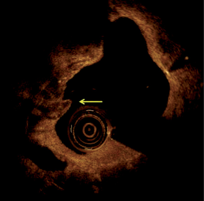
Figure 1. Optical coherence tomography in the RCA revealing calcified tissue with a sharp calcium spicule protruding into the lumen (arrow).

Conflict of interest statement

The authors have no conflicts of interest to declare.
Volume 10 Number 5
Sep 28, 2014
Volume 10 Number 5
View full issue


Key metrics

Suggested by Cory

Image – Interventional flashlight

10.4244/EIJ-D-20-00507 Aug 6, 2021
Optical coherence tomography and tailored treatment of in-stent restenosis
Jia H et al
free

Research Correspondence

10.4244/EIJ-D-24-01113 Jul 7, 2025
Restenosis patterns after percutaneous coronary intervention with drug-coated balloons for de novo coronary lesions
Matsuda K et al
free

10.4244/EIJV7I3A65 Jul 29, 2011
How should I treatsevere calcific coronary artery disease?
Fernandez J et al
free

Viewpoint

10.4244/EIJ-D-25-01066 May 4, 2026
Intracoronary imaging guidance for de novo coronary lesion treatment with drug-coated balloons
Amabile N et al
free

Image – Interventional flashlight

10.4244/EIJ-D-19-00268 Jul 17, 2020
Intravascular lithotripsy for recurrent restenosis caused by severe calcific neoatherosclerosis
Salazar C et al
free

Clinical Research

10.4244/EIJ-D-21-00504 Mar 18, 2022
Prevalence, predictors, and outcomes of in-stent restenosis with calcified nodules
Tada T et al
free
Trending articles
334.7

State-of-the-Art Review

10.4244/EIJ-D-21-00904 Apr 1, 2022
Antiplatelet therapy after percutaneous coronary intervention
Angiolillo D et al
free
84.05

State-of-the-Art

10.4244/EIJ-D-23-00840 Sep 2, 2024
Aortic regurgitation: from mechanisms to management
Baumbach A et al
free
45.75

State-of-the-Art

10.4244/EIJ-D-25-01346 May 4, 2026
Slow flow and no reflow after percutaneous coronary intervention
Brugaletta S et al
free
42.15

State-of-the-Art

10.4244/EIJ-D-25-00896 Apr 6, 2026
Pretreatment with antiplatelet agents in patients undergoing coronary revascularisation
Kaur G et al
free
38.2

State-of-the-Art

10.4244/EIJ-D-24-00195 Apr 7, 2025
Percutaneous coronary intervention for calcified and resistant lesions
Pesarini G et al
free
33.5

Original Research

10.4244/EIJ-D-26-00032 May 15, 2026
Glucocorticoids to reduce permanent pacemaker implantation after TAVI: the GLUCO-TAVI randomised trial
Fuertes-Kenneally L et al
32.2

State-of-the-Art

10.4244/EIJ-D-25-00874 Jun 1, 2026
TAVI and coronary interventions: indications, technical considerations, and clinical scenarios
Aquino Bruno H et al
free
29.2

Original Research

10.4244/EIJ-D-25-01370 May 21, 2026
Prognostic value of early haemodynamic valve deterioration after TAVI
Trimaille A et al
29.2

Original Research

10.4244/EIJ-D-25-01370 Jun 1, 2026
Prognostic value of early haemodynamic valve deterioration after TAVI
Trimaille A et al
X

PCR
Impact factor: 9.2
2025 Journal Citation Reports®
Science Edition (Clarivate Analytics, 2026)
Online ISSN 1969-6213 - Print ISSN 1774-024X
© 2005-2026 Europa Group - All rights reserved