A 50-year-old diabetic man underwent percutaneous coronary intervention of an LAD/diagonal bifurcation lesion with deployment of a 3.0×28 mm Absorb bioresorbable vascular scaffold (Abbott Vascular, Santa Clara, CA, USA) in the LAD (Online Figure 1A, Moving image 1). After through-the-strut dilation, a 2.5×38 mm XIENCE PRIME™ stent (Abbott Vascular) was deployed in the side branch with a 3.0×15 mm non-compliant balloon positioned in the LAD. A final kissing balloon inflation (FKB) was then performed at 4 atmospheres (Online Figure 1B, Moving image 2). Optical coherence tomography 3-D reconstruction revealed preserved integrity of the bioresorbable vascular scaffold (BVS) rings and complete opening of the side branch ostium (Figure 1, Online Figures 2-5, Moving image 3-5). Inflation through the struts of the BVS is feasible but must be performed slowly. Aggressive inflation can result in ring fracture, instability of the scaffold, and potentially thrombosis. BVS disruption has been observed clinically, and in bench testing after FKB at or just above the maximal recommended diameter for the BVS, using Finet’s principle. If FKB is necessary, inflation must be performed slowly and to low pressures only, maintaining the combined inflated balloon diameter below the recommended limit of the device. Intravascular imaging of the final result is mandatory.
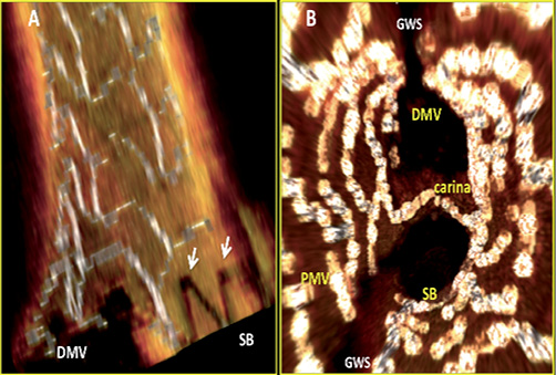
Figure 1. A) 3-D OCT of the BVS/EES interface at the side branch ostium, B) fly-through view from the side branch. DMV: distal main vessel; GWS: guidewire shadow; PMV: proximal main vessel; SB: side branch. Arrows indicate XIENCE V stent struts
Guest Editor
This paper was Guest Edited by Carlo Di Mario, MD, PhD; NHLI Cardiovascular Biomedical Research Unit, Royal Brompton Hospital, London, United Kingdom.
Conflict of interest statement
V. Džavík has received educational and travel grants, and speaker honoraria from Abbott Vascular, and educational grants from St. Jude Medical. The other authors have no conflicts of interest to declare.
Online data supplement
Online Figure 1. LAD diagonal bifurcation before (A) and after (B) T-stenting.
Online Figure 2. 2-D OCT images through the side branch showing BVS/EES overlap at the carina (A), the carina (B) and proximal to the carina (C).
Online Figure 3. 3-D OCT reconstruction of the side branch ostium.
Online Figure 4. 3-D OCT details of the BVS/EES interface at the side branch ostium.
Online Figure 5. Fly-through views from the distal main vessel and from the side-branch.
Moving image 1. Pre-PCI angiogram of a Medina 0,1,1 LAD/D1 bifurcation lesion.
Moving image 2. Final post-PCI angiogram.
Moving image 3. An OCT pullback from the diagonal branch to the proximal LAD.
Moving image 4. An OCT pullback from the mid LAD to the proximal LAD.
Supplementary data
To read the full content of this article, please download the PDF.
Moving image 1. Pre-PCI angiogram of a Medina 0,1,1 LAD/D1 bifurcation lesion.
Moving image 2. Final post-PCI angiogram.
Moving image 3. An OCT pullback from the diagonal branch to the proximal LAD.
Moving image 4. An OCT pullback from the mid LAD to the proximal LAD.