A 55-year-old male underwent an exercise treadmill test which was positive at stage 1.
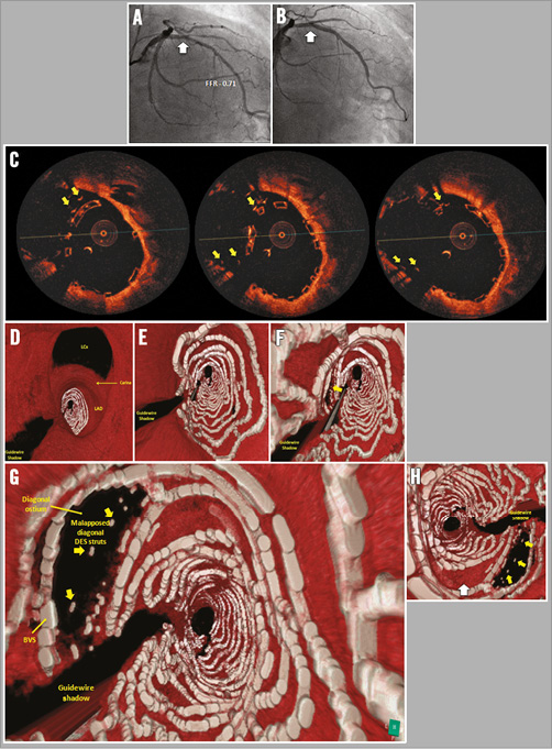
Coronary angiography (Figure 1A, Moving image 1) demonstrated a long segment of disease in the proximal-mid left anterior descending (LAD) artery. A fractional flow reserve study was positive at 0.71, with no localising lesion on hyperaemic pullback. In addition, >5 mm disease was present in the ostial-proximal first diagonal. A hybrid mini-crush approach was adopted.
The LAD was predilated with a 3.0 mm non-compliant (NC) balloon. A 2.25×24 mm Promus PREMIER (Boston Scientific, Marlborough, MA, USA) drug-eluting stent (DES) was implanted in the diagonal and three overlapping Absorb bioresorbable vascular scaffolds (BVS) (3.5×28 mm, 3.5×28 mm, 3.5×18 mm) (Abbott Vascular, Santa Clara, CA, USA) in the LAD. Post-dilatation of the LAD was performed with a 3.75 mm NC balloon. Low-pressure (six atmospheres) mini-kissing balloon post-dilatation (KBPD) was then performed using 3.0 mm and 2.5 mm NC balloons in the main and side branch (SB), respectively. The final angiographic result was excellent (Figure 1B, Moving image 2). Optical coherence tomography (OCT) was then performed using a Dragonfly™ Duo catheter (St. Jude Medical, St. Paul, MN, USA), and showed an acceptable result (Figure 1C).
Offline, fly-through three-dimensional reconstruction of the OCT images showed incomplete apposition of the DES at the diagonal ostium (Figure 1D-Figure 1G), in addition to underexpansion of the BVS in the proximal segment (Figure 1H, white arrow).

Figure 1. Two- and three-dimensional OCT of a hybrid mini-crush approach to LAD-diagonal bifurcation. A) & B) Pre- and post-coronary angiography. C) Corresponding 2D OCT. D-G) Fly-through 3D OCT from left main to LAD-diagonal. H) Reverse view LAD-diagonal bifurcation.
To ensure adequate apposition of the BVS and the SB stent, we propose higher pressure sequential balloon inflation in the BVS and the SB, followed by low-pressure mini-KBPD to correct any resulting distortion. The risk of BVS strut fracture using a 2.5 mm NC balloon for SB dilatation is ~13%. This, perhaps, could be mitigated by slower balloon inflation and/or using a smaller balloon at higher pressure.
Conflict of interest statement
The authors have no conflicts of interest to declare.
Supplementary data
Moving image 1. Coronary angiography demonstrating disease in the proximal-mid LAD and ostial-proximal first diagonal.
Moving image 2. Coronary angiography demonstrating final angiographic result.
Supplementary data
To read the full content of this article, please download the PDF.
Moving image 1. Coronary angiography demonstrating disease in the proximal-mid LAD and ostial-proximal first diagonal.
Moving image 2. Coronary angiography demonstrating final angiographic result.

