Abstract
Recommended techniques for bifurcation stenting continue to be revised with specific attention to bioresorbable scaffolds (BRS). Optimal procedural success and long-term outcomes with BRS can perhaps be improved with careful attention to implantation techniques. Good vessel preparation is imperative for optimal expansion of the scaffold, and proper vessel sizing is necessary to ensure compliance with scaffold expansion limits and preservation of proper scaffold function. The European Bifurcation Club (EBC) recommends provisional stenting for the majority of bifurcation lesions: permanent metallic stents are sized according to the distal vessel diameter, with subsequent post-dilatation of the proximal vessel to ensure stent apposition in the proximal main vessel. Recent BRS-specific modifications to the EBC recommendation suggest that selecting the scaffold size based on the diameter of the proximal main vessel can mitigate the risk of overexpansion and potential strut fracture. Expansion of the BRS requires a thoughtful balance between the risk of malapposition associated with underdeployment and the risk of strut fracture due to overdeployment. Post-dilatation of scaffolds should be performed, always respecting the maximum expansion limit, to correct any potential scaffold malapposition and minimise flow disturbances. Finally, dual antiplatelet therapy plays an important role in BRS bifurcation treatment to avoid thromboembolic events.
Introduction
Bifurcation lesions are an important part of routine percutaneous coronary intervention. Single-arm bioresorbable scaffold (BRS) trials have reported a large proportion of cases involving bifurcations. Considerable effort has gone into developing techniques to optimise procedural success and long-term outcomes when using permanent metallic stents1. These efforts have concentrated on minimising stent material (excess metal) while maximising flow quality (maximising volume and minimising variations in the local haemodynamic environment that lead to wall shear stress), always allowing for the permanent nature of the implant(s) (e.g., minimising the permanent nature of side branch jailing) and assuming that acute angiographic success will yield good long-term results. The advent of bioresorbable scaffolds (BRS) adds new possibilities for the treatment of bifurcation lesions. Traditional recommendations for metallic stents can be re-examined to take into consideration the differences between metallic and resorbable platforms, whilst also considering the temporal evolution of the treatment site as the implant(s) resorbs. In this article, we review these differences and take into account the current limitations of resorbable platforms in the context of bifurcation lesion treatment. Successful outcomes and low complication rates for BRS bifurcation stenting can be ensured with careful attention to implantation techniques.
Lesion preparation
Bifurcations are, by their very nature, a nidus for flow disturbance that can cascade into mechanisms that lead to obstructive plaque formation. Plaque content in bifurcations is of a complexity equal to or greater than plaque found in most locations of the coronary tree. In particular, the incidence of calcifications and/or fibrosis is high in bifurcations. Calcification can present a challenge for device crossing, particularly with BRS, where the crossing profile is still similar to that of first-generation metallic devices. Predilation in these lesions becomes critical, not only to ensure that the BRS can cross the lesion, but also to anticipate any fibrosis that may lead to inadequate expansion of the scaffold. The use of a non-compliant predilation balloon with a diameter equal to the reference vessel diameter, and techniques such as rotablation and/or scoring balloons can help prepare these lesions to ensure uniform, predictable, and optimal expansion of the scaffold, and good acute lumen gain. Achieving a residual stenosis less than 20-40% is recommended by the manufacturers of currently approved products. These techniques have been applied with excellent acute success2.
Vessel sizing, implantation and post-dilatation
Following appropriate lesion preparation, acute procedural success is contingent upon optimal scaffold sizing prior to scaffold placement. BRS sizing and subsequent expansion require a thoughtful balance between the risk of malapposition associated with underdeployment and the risk of strut fracture due to overdeployment (exceeding scaffold expansion limits). BRS manufacturers provide product-specific recommendations for the maximum expansion possible for each nominal scaffold diameter. At present, both commercially available products, Absorb BVS (Abbott Vascular, Santa Clara, CA, USA) and DESolve (Elixir Medical Corp., Sunnyvale, CA, USA), state a maximum functional diameter limit of 0.5 mm above the labelled nominal diameter. These maximum expansion diameters must be respected, since proper scaffold functionality (including structural integrity, acute radial strength, luminal support time, etc.) is only possible at or below this diameter. Expansion beyond these limits can potentially result in strut fracture or in deterioration of scaffold mechanical properties that are critical to long-term device function/arterial segment healing and outcomes, even in the absence of fracture.
The European Bifurcation Club (EBC) recommends provisional stenting as the preferred technique for the majority of bifurcation lesions. In this technique, the main vessel is stented first and the side branch (SB) is only stented in case of severe stenosis or flow limitations to the side branch after main vessel stenting3. This provisional approach remains the default bifurcation technique for BRS as well, but scaffold size selection is more operator- and anatomy-dependent. As per the manufacturers’ recommendations, provisional bifurcation treatment can be performed by placing BRS directly across SBs less than or equal to 2 mm. Enrolment criteria in all BRS trials have allowed for such practices, and shown an elevated rate of non-Q-wave myocardial infarction (NQMI), but without detrimental acute or long-term consequences. Muramatsu et al concluded that the elevated rate of NQMI might be due to the larger footprint of BRS, leading to a higher rate of side branch blockages compared to DES implantation4. Nonetheless, SB flow should be preserved, especially in larger SBs. Fenestration, or dilation through a scaffold structural cell into an SB, should be performed only when SB flow is compromised, using the minimal balloon diameter possible. Larger diameter balloons may disrupt the scaffold cell integrity, with higher angulations and balloon diameters disrupting ring integrity at higher rates.
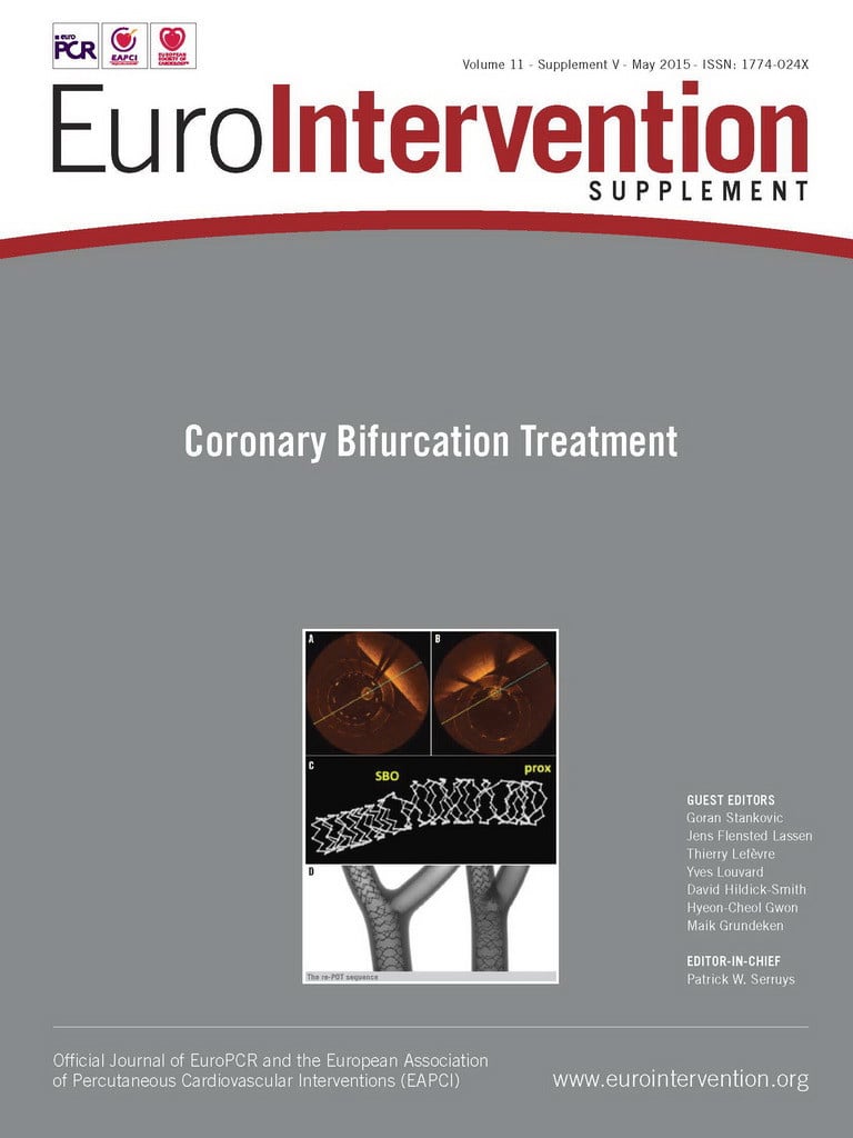
According to the EBC recommendation, permanent metallic stents should be sized according to the distal diameter, with subsequent post-dilatation of the proximal vessel segment with a larger balloon to ensure stent apposition in the proximal main vessel (also referred to as “proximal optimisation technique” or POT). This technique may be used with BRS when the diameter of the proximal main vessel is smaller than the scaffold expansion limit. The initial scaffold deployment to the diameter of the distal segment leaves the deployed BRS undersized and malapposed in the segment proximal to the bifurcation. Low-pressure POT with a non-compliant (NC) balloon can correct this proximal malapposition without oversizing the distal scaffold, thereby preserving the BRS integrity5.
More recent modifications of the EBC recommendation, specific to BRS, suggest that selecting the scaffold size based on the diameter of the proximal main vessel can mitigate the risk of BRS overexpansion and potential strut fracture5,6. Deploying the scaffold at low pressure avoids damaging the distal main vessel, with subsequent POT with a short NC balloon, sized for the proximal main vessel, inflated at high pressure to ensure apposition at the proximal segment. An important consideration for this latter approach is the post-dilatation diameter of the proximal MV, which should not exceed the expansion limit for the BRS at the desired inflation pressures. Similar attention should be paid to the scaffold’s expansion in the distal segment of the vessel, which should meet the nominal deployment diameter in order to ensure proper scaffold function.
Routine final “kissing” balloon dilatation (FKBD) is not recommended with BRS due to the risk of oversizing, i.e., the combined diameters of the two balloons may exceed the scaffold expansion limits and result in ring fracture. Recently, mini-FKBD (or “snuggle” balloon dilatation) was proposed, with low-pressure inflation of minimally overlapped NC balloons5,7,8. At low pressure, the scaffold constrains the balloon expansion. It has been suggested that higher pressures increase the risk of ring fracture because the scaffold can no longer constrain the balloon expansion, and the scaffold expansion limit is exceeded3,5,7. While FKBD performed at low inflation pressures might be important when a two-stent bifurcation technique is used, sequential inflations with NC balloons are preferable in a provisional single-stent procedure to optimise BRS apposition. As described above, if the SB is compromised, a scaffold cell may be opened with an undersized NC balloon and POT performed with a larger balloon in the proximal MV to correct the typical scaffold malapposition often observed opposite the SB take-off.
Pre-implantation QCA, as well as intravascular imaging, such as IVUS and OCT, pre- and post-implantation, can be used to ensure optimal sizing of the BRS and to confirm that the scaffold is well apposed and strut integrity is maintained after the bifurcation procedure. Mathey et al9 investigated the safety and efficacy of the Absorb BVS in a real-world setting and demonstrated that, despite visual overestimation of baseline reference vessel diameter (RVD) compared to QCA, the final minimal lumen diameter (MLD) closely matched the baseline RVD. Visual estimation may be acceptable for scaffold sizing, but disrupted scaffold struts cannot be visualised by angiography. Dzavík et al10 recommend intravascular imaging, preferably with OCT or IVUS, whenever dilating BRS struts, performing POT, FKBD, or deploying two BRS, to ensure scaffold integrity in the final result. Extensive use of post-dilatation, within scaffold expansion limits, has been shown, with limited sample size, to achieve excellent acute and midterm success5.
Beyond post-dilation
The integrity of scaffold rings is important to the acute performance of the BRS, as radial strength is provided by the circumferentially oriented ring elements of the structure, and is linearly dependent on the number of integral rings in the structure. Preserving ring integrity thus ensures sufficient radial strength to support the lumen. Scaffold links are oriented along the axis of the vessel and therefore provide uniform distribution of the rings during delivery and deployment, but have no role in luminal support.
During SB dilatation, a scaffold cell is opened, and fractures have been shown to occur at similar rates in both the rings and links for certain larger balloon sizes and more severe SB angulations5. The degree of cell opening is highly dependent upon the configuration of the scaffold: a scaffold deployed into a well prepared, straight vessel will have uniform cell opening, whereas a scaffold deployed in severe SB angulations will have variable fenestrations depending on the degree of curvature and the cell’s position along the convex or concave side of the curve. The fractures identified in the Ormiston study5 occurred in single struts and were not associated with strut malapposition. At high pressures during mini kissing balloon post-dilatation (mini-KBPD), multiple ring fractures could occur, with the ends of fractured struts malapposed, overlapping and projecting into the lumen, creating a potential nidus for adverse clinical events5. When the techniques described here were followed, namely careful BRS size selection and respect for expansion limits, scaffold integrity was maintained.
Improper apposition of the BRS in the main branch can jail the SB and give rise to a more aggressive “Darcy friction”-derived pressure drop across the jailed SB (along the main vessel). The consequence of an increased pressure drop across the jailed SB is twofold. First, there is a reduction of flow in the MB, which may result in insufficient perfusion/ischaemia in the myocardium distal to the side branch. Second, normal and shear stresses due to the malapposition, or simply the thicker struts associated with commercial BRS, can contribute to skin friction on the fluid and platelets. These stresses on the platelet surface can activate a biochemical signal cascade in the platelet, resulting in secretion of ADP, serotonin, and other granule chemicals. Platelet activation, in combination with low flow rates in the side branch, may lead to thromboembolic events.
A recent analysis comparing shear stress patterns as a function of strut thickness and/or strut malapposition has demonstrated that malapposition plays a much larger role in shear stresses than does strut thickness11. Thus, correcting malapposition, and mitigating the associated flow compromise in the SB, is a critical part of the procedure, and will remain so despite the development of next-generation scaffolds with thinner struts.
More complex anatomy requiring a priori two scaffold approaches should be considered carefully, and most likely under investigational conditions, to monitor the efficacy of results over the long term12. With current-generation scaffolds, some physicians may choose to use thinner-strutted metallic stents in side branches to preserve the luminal diameter and flow volume, and techniques with least strut layering should be preferred (i.e., T, T and small protrusion [TAP]) when a second stent or scaffold is needed.
Antiplatelet therapy
As natural sites of flow disturbance, including slow recirculation and high flow/shear rates, bifurcations can present their own challenges. Implantation of stents and/or scaffolds to improve flow in the vessel(s) can result in further alteration in the boundary layer flow patterns, an effect that is much more pronounced at bifurcations. Thus, proper anticoagulation is even more important in bifurcation treatment. In addition, when extensive vessel preparation has been performed, the combined effect of vessel wall injury and implantation of scaffolds can leave the segment particularly prone to platelet adhesion and activation. More potent antiplatelet therapy in such cases has been reported, and should be considered.
Conclusion
In summary, bifurcation treatment with BRS has been performed successfully with a few considerations specific to the limitations of BRS. Proper sizing of the vessel is necessary to ensure that scaffold expansion limits are observed and that all aspects of proper scaffold function are preserved. Uniform, predictable, and optimal expansion of the scaffold is more likely with proper vessel preparation. Post-dilatation of scaffolds should be done according to EBC recommendations, always respecting the maximum expansion on the label. Finally, flow disturbances should be minimised and compromised SB flow eliminated prior to concluding the procedure. Dual antiplatelet therapy plays an even more important role in bifurcation treatment to avoid thromboembolic events.
Conflict of interest statement
J. Fox, S. Hossainy and R. Rapoza are current full-time employees of Abbott Vascular. P. Serruys has no conflicts of interest to declare.

