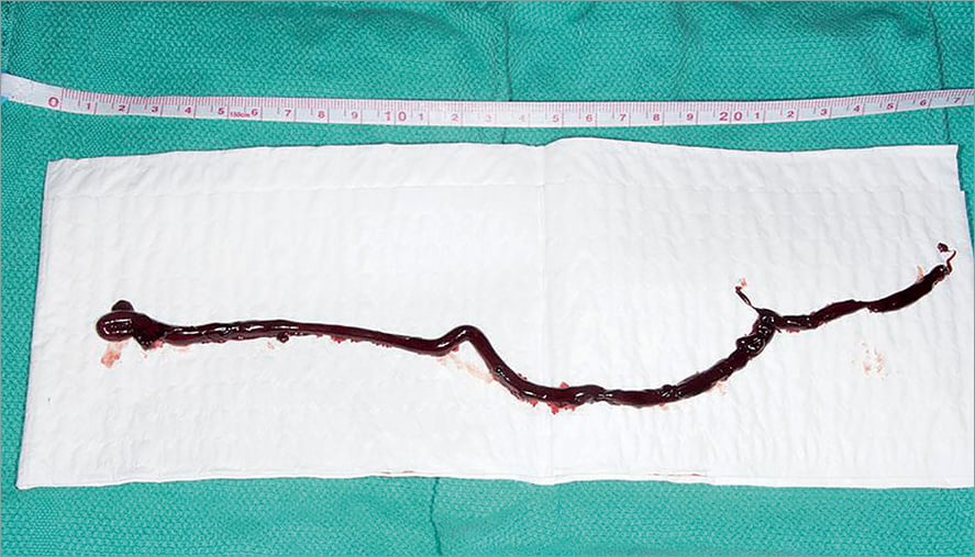

Figure 1. Picture of the catheter-related thrombus (measuring at least 27 cm), which remained almost completely intact despite the aspiration process.
A 90-year-old man, with a past medical history significant for type 2 diabetes mellitus, atrial fibrillation, chronic lymphocytic leukaemia, and prior sutureless aortic valve replacement with a Perceval valve (Sorin Group, Saluggia, Italy), was admitted to our institution because of decompensated heart failure caused by severe degenerative mitral regurgitation (MR). The Heart Team deemed the patient to be at very high risk for a reoperative open heart surgery. Hence, he was referred for a MitraClip® (Abbott Vascular, Santa Clara, CA, USA) procedure. The MR was significantly reduced after the deployment of one clip. The 24 Fr steerable guide catheter was thus removed from the right femoral vein. Despite appropriate flushing of the guide catheter and adequate anticoagulation with intravenous heparin during the entire MitraClip procedure (ACT >250 sec), a giant, very mobile, moulding thrombus of the steerable guide catheter was seen in the right atrium on transoesophageal echocardiography (Moving image 1). Through a long 14 Fr sheath placed in the left femoral vein, we were able to aspirate the 27 cm catheter-related “snake-like” thrombus completely (Figure 1). The patient was rapidly extubated. A ventilation perfusion scan did not reveal any signs of pulmonary embolism. The patient was put on IV heparin after the procedure and then bridged to Coumadin. He was discharged home three days later. Thrombi forming within large catheters used for structural heart interventions are usually multicausal, can have catastrophic consequences and should be avoided by constantly flushing the catheters as well as by aiming for a very high level of anticoagulation during the procedure. The optimal treatment remains controversial, including anticoagulation, thrombolysis, surgical thrombectomy and percutaneous thromboaspiration1.
Conflict of interest statement
J. Rodés-Cabau has received research grants from Edwards Lifesciences, Medtronic and Heart Leaflet Technologies. The other authors have no conflicts of interest to declare.
Supplementary data
Moving image 1. Transoesophageal echocardiogram showing a giant, very mobile thrombus located in the right atrium.
Supplementary data
To read the full content of this article, please download the PDF.
Moving image 1. Transoesophageal echocardiogram showing a giant, very mobile thrombus located in the right atrium.

