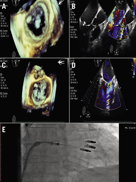

Figure 1. Transoesophageal echocardiography. A) Preprocedural 3D: a large vegetation of 1.5 by 1.7 cm at the free edge of the anterior mitral leaflet (A2). B) Preprocedural 2D: torrential mitral regurgitation with no coaptation of the leaflets. C) Post-procedural 3D: reduction of MR to 1+. D) Post-procedural 2D: reduction of MR to 1+. E) The three clips in the mitral valve.
The usage of MitraClip® devices (Abbott Vascular, Santa Clara, CA, USA) for treating mitral regurgitation due to endocarditis has been reported previously1. Six cases of infective endocarditis (IE) after repair with the MitraClip have also been reported. Three of the six cases were caused by staphylococcus aureus and two of these patients died. Most of the patients were treated surgically2.
An 82-year-old male patient with a history of deteriorating heart failure was admitted to our hospital. His past medical history revealed chronic liver cirrhosis due to hepatitis C, chronic renal failure, myelodysplastic syndrome and severe cachexia. Two years ago, he was evaluated for a persistent febrile state without any conclusive diagnosis.
Transoesophageal echocardiography (TEE) revealed torrential mitral regurgitation with no coaptation of the leaflets and a large vegetation with dimensions 1.5-1.7 cm at the free edge of the anterior mitral leaflet (A2) (Figure 1A, Figure 1B, Moving image 1, Moving image 2). Evaluation for possible active endocarditis was negative. After careful evaluation by our Heart Team, the patient was not offered the option of operation because of his extreme high risk (his logistic EuroSCORE was 48%).
Finally, a percutaneous mitral valve repair was performed using the MitraClip device as a rescue/palliative procedure. Three clips were implanted side by side in the A2-P2 area trapping the large vegetation and reducing MR to 1+ (Figure 1C-Figure 1E, Moving image 3-Moving image 5). The patient had an uneventful recovery.
Percutaneous repair of the mitral valve using the MitraClip device is feasible even in the setting of a large vegetation in the grasping zone. This option might be considered as a valuable alternative in extremely high-risk patients. To the best of our knowledge, this is the first case that has been performed in this setting.
Conflict of interest statement
The authors have no conflicts of interest to declare.
Supplementary data
Moving image 1. Preprocedural 3D TEE. A large vegetation at the free edge of the anterior mitral leaflet (A2).
Moving image 2. Preprocedural 2D TEE. Torrential mitral regurgitation with no coaptation of the leaflets.
Moving image 3. Post-procedural 3D TEE. MR 1+.
Moving image 4. Post-procedural 2D TEE. MR 1+.
Moving image 5. Three clips in mitral valve.
Supplementary data
To read the full content of this article, please download the PDF.
Moving image 1. Preprocedural 3D TEE. A large vegetation at the free edge of the anterior mitral leaflet (A2).
Moving image 2. Preprocedural 2D TEE. Torrential mitral regurgitation with no coaptation of the leaflets.
Moving image 3. Post-procedural 3D TEE. MR 1+.
Moving image 4. Post-procedural 2D TEE. MR 1+.
Moving image 5. Three clips in mitral valve.

