Optical Coherence Tomography (OCT) is establishing itself as the new standard for in vivo assessment of stent coverage and strut apposition.1-3 This edition of EuroIntervention contains two studies that illustrate the evolving role of OCT in interventional cardiology.
Capodanno and collaborators report a comparison between OCT versus intravascular ultrasound (IVUS) in assessing stented vessels4. They observe a good correlation between the two modalities with respect to morphometric measurements. OCT detects more neointimal hyperplasia (NIH) than IVUS, which is likely due to resolution differences between methods5,6. The ability of OCT to render near histology-level NIH detection and quantification has been recently confirmed in a well conducted pre-clinical study.7 The observed differences in NIH quantification in Capodanno’s study would likely be amplified if more potent DES were examined. Indeed, we have observed a greater discrepancy between OCT and IVUS in sirolimus and paclitaxel eluting stents as opposed to bare metal stents1.
The authors avoided using the term “NIH” and instead adopt “tissue coverage area” (TCA). By abandoning the term NIH, the authors herald a new era in intravascular imaging. “NIH” was coined decades ago at the advent of the balloon angioplasty era where one might expect a more predictable vascular response to mechanical injury characterised by predominantly smooth muscle cell proliferation and extra-cellular matrix elaboration.8 However, in the modern era of complex drug, polymer and vessel interactions, a more inclusive approach must be applied to better understand the vascular response to injury and tissue in-growth after stent placement.
It is important for the reader to understand the differences between cross-section level analysis and strut-level analysis. OCT quantification of tissue growth at a cross-sectional level, while albeit more sensitive, is similar in principles to conventional IVUS quantification. But we can afford to look much closer and in more details with OCT imaging, and TCA should not be mistakenly interpreted as “strut-level analysis” for stent coverage. “Strut coverage” represents a qualitative assessment on the presence of tissue overlaying each strut. This tissue can also be quantified as strut-level neointimal thickness. Strut-level coverage analysis was also performed in the study, and a good inter-observer concordance was demonstrated, but this was not central to the author’s findings. However, strut-level analysis is rapidly being adopted as a surrogate endpoint in clinical trials of DES. We now realise that a single cross-section may house struts with strikingly different quality and quantity of tissue coverage1. This heterogeneous response may only be discriminated by means of strut-level analysis.
Beyond strut-level analysis, tissue characterisation is the next frontier for intracoronary OCT. Current OCT technologies cannot differentiate histological properties of the vasculature, and early efforts have used the broader term “abnormal intraluminal tissue (AIT)”2 to distinguish normal- from ill-appearing tissue growth based upon properties of scattering and absorption of light in targeted tissues. Similarly, a “restenotic tissue classification” has been proposed9. However, the technology and its imaging post-processing will need to evolve further. Serial imaging studies and histopathological validation will be required to achieve a better understanding of the host response to different stent types and for translation of these findings into clinical practice.
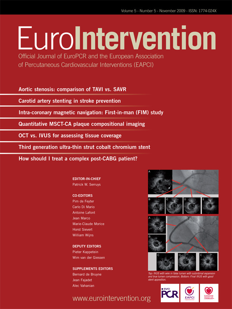
The report of Tyczynski and collaborators illustrates how OCT can provide a new perspective on “old” questions10. Bifurcation coronary artery disease frequently plagues interventional cardiologists, and optimal management strategies continue to evolve. The fact that a 9-patient study made it to press, underscores the impact of this novel surrogate imaging method to our field. Indeed, the bifurcation lesion represents one of the most attractive clinical applications of OCT. The authors found a higher number of “malapposed” struts at the level of the ostium of the side branch, despite the use of a dedicated bifurcation stent and appropriate final kissing-balloon inflation. In retrospect, one might ask if this study discourages the use of dedicated bifurcation stents.... We don’t think so, as having more “malapposed” struts towards the ostium is expected even if single stent approaches are used. We have previously assessed 6-month outcomes of DES struts “jailing” side branches using OCT in 61 coronary bifurcations treated with single stent techniques without kissing-balloon inflation11. The quantity and quality of tissue coverage, as well as strut apposition, varied widely among stent types and, most importantly, within the same stent cross-section depending upon whether the struts were located at the ostium, adjacent to the ostium, or opposite to the side branch11.
Assessment and quantification of stented bifurcations still needs further refinement, and the potential application of 3D imaging to properly depict anatomical intricacies holds promise. The new frequency domain OCT systems, such as the commercially available C7 XR (LightLab Imaging Inc., Westford, MA, USA), has a much higher sampling rate and faster pullback speed (20 mm/sec), which is expected to minimise the effect of the cardiac movement allowing more accurate 3D image reconstructions.
OCT’s ability to image embryonic hearts12, preclinical small and large animals and human coronaries, provides clinician/scientists a valuable translational research tool. Furthermore, OCT’s in vivo serial imaging capabilities offers an opportunity to assess the temporal evolution of atherosclerosis and arterial healing, expanding considerably our current knowledge base in a way distinct from the traditional single snapshots derived from necropsy specimens.
Vascular healing is a complicated and dynamic process which may not develop in a linear fashion as one may think. Obviously, histology will continue to play an important role in device testing, but it also needs to mature as a technology. Novel experimental techniques that enable functional assessment of the endothelium will permit investigators to better dissect the differences between an intact endothelial monolayer that is dysfunctional, and potentially prothrombotic from one which promotes vascular health and homoeostasis. Similar techniques may also permit in situ 3D reconstruction of the vessel13. Together with OCT imaging, these contemporary histologic techniques will help us reappraise unanswered “old” questions. Nevertheless, some premises are still the same. Only well conducted studies with rigorous scientific methodology will transform these nice pictures into relevant clinical findings.