IMAGE IN CARDIOLOGY

DOI: 10.4244/EIJ-D-16-00412

Incidental finding of a bubble-shaped mass during coronary angiography

Tomasz Bochenek*, MD, PhD; Błażej Kusz, MD; Michał Lelek, MD, PhD; Marek Grabka, MD, PhD; Katarzyna Mizia-Stec, MD, PhD

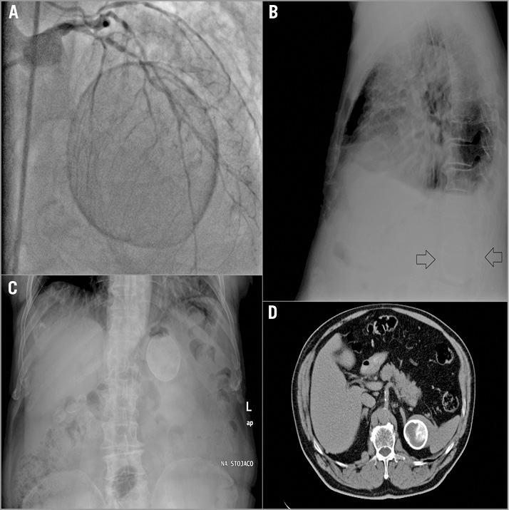
An 84-year-old man with a history of hypertension, smoking, and diabetes mellitus was admitted to our institution with non-ST-elevation myocardial infarction. Coronary angiography revealed multivessel coronary disease. To our surprise, a large bubble-shaped mass was found in projection LAO 7, CAU 36 (Panel A). We performed a chest X-ray that visualised the presence of a mass lying under the diaphragm (Panel B). An abdominal X-ray revealed a calcified shaded area measuring ca. 60 mm in the left supra-abdominal space (Panel C). Subsequently, abdominal ultrasonography was carried out; no abnormalities were shown. Finally, computed tomography of the abdomen confirmed that the mass was a calcified cyst of the spleen, most probably idiopathic (Panel D). Following the Heart Team’s decision, the patient underwent successful percutaneous coronary intervention of the left anterior descending artery and was discharged four days later. This case presents an unusual scan of a routine coronary angiography and illustrates the importance of a careful interpretation of every fluoroscopic image that is used in contemporary medicine.

Conflict of interest statement

The authors have no conflicts of interest to declare.

Volume 12 Number 16
Mar 20, 2017
Volume 12 Number 16
View full issue


Key metrics

Suggested by Cory

IMAGE IN CARDIOLOGY

10.4244/EIJV11I4A86 Aug 20, 2015
Calcified nodule: a double paradox on coronary imaging
Alfonso F et al
free

Debate

10.4244/EIJ-E-23-00002 Jun 5, 2023
All calcified nodules are made equal and require the same approach: pros and cons
Guagliumi G et al
free

IMAGE IN CARDIOLOGY

10.4244/EIJV10I5A104 Sep 28, 2014
High definition OCT image of sharp calcium spicule
Garrido E et al
free

10.4244/EIJV10I4A91 Aug 19, 2014
How should I treat an undeployed stent stuck in the proximal LAD? Going for another round...
Locca D and Eeckhout E
free

10.4244/EIJV16I5A65 Aug 28, 2020
OCT-derived coronary calcified nodules as a predictor of high-risk patients
Akasaka T and Kubo T
free
Trending articles
200.45

State-of-the-Art

10.4244/EIJ-D-21-00089 Jun 11, 2021
Intracoronary optical coherence tomography: state of the art and future directions
Ali ZA et al
free
154.43

State-of-the-Art

10.4244/EIJ-D-22-00776 Apr 3, 2023
Computed tomographic angiography in coronary artery disease
Serruys PW et al
free
92.95

State-of-the-Art Review

10.4244/EIJ-D-20-01296 Aug 27, 2021
Management of cardiogenic shock
Thiele H et al
free
47.4

State-of-the-Art

10.4244/EIJ-D-24-00386 Feb 3, 2025
Mechanical circulatory support for complex, high-risk percutaneous coronary intervention
Ferro E et al
free
43.65

Clinical research

10.4244/EIJ-D-23-00590 Dec 4, 2023
Prognostic impact of cardiac damage staging classification in each aortic stenosis subtype undergoing TAVI
Nakase M et al
free
36.5

State-of-the-Art

10.4244/EIJ-D-23-00448 Jan 15, 2024
Coronary spasm and vasomotor dysfunction as a cause of MINOCA
Yaker ZS et al
free
34.75

State-of-the-Art

10.4244/EIJ-D-23-00606 Jan 1, 2024
Targeting inflammation in atherosclerosis: overview, strategy and directions
Waksman R et al
free
X

The Official Journal of EuroPCR and the European Association of Percutaneous Cardiovascular Interventions (EAPCI)

EuroPCR EAPCI
PCR ESC
Impact factor: 7.6
2023 Journal Citation Reports®
Science Edition (Clarivate Analytics, 2024)
Online ISSN 1969-6213 - Print ISSN 1774-024X
© 2005-2025 Europa Group - All rights reserved