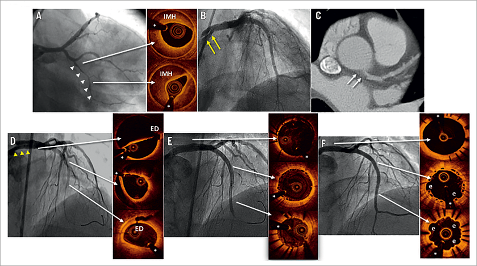

Figure 1. Angiographic, OCT and coronary CT findings. A) SCAD at distal LCX (white arrowheads). OCT confirmed a long IMH. B) ICAD at LM (yellow arrows). C) Progression of the LM ICAD on coronary CT (white arrows). D) Extension of the ICAD involving LAD with distal occlusion (yellow arrowheads). OCT runs with multiple ED. E) Excellent final angiographic and OCT result after DES and BVS implantation. F) Persistent good angiographic and OCT result on follow-up.
A 47-year-old woman was admitted with an acute coronary syndrome. Coronary angiography revealed lumen narrowing at the distal left circumflex (LCX) (Panel A, white arrowheads). Optical coherence tomography (OCT) confirmed the presence of a spontaneous coronary artery dissection (SCAD) with a long intramural haematoma (IMH). A final projection disclosed a confined iatrogenic coronary artery dissection (ICAD) induced by the guiding catheter at the left main (LM) (Panel B, yellow arrows). At this point, a conservative strategy was selected. Despite an unremarkable initial clinical course, a scheduled coronary CT suggested progression of the LM ICAD (Panel C, white arrows), prompting a decision for intervention. The coronary angiogram confirmed the extension of the ICAD to the proximal and mid left anterior descending coronary artery (LAD) with distal occlusion (Panel D, yellow arrowheads). OCT depicted multiple entry doors (ED) at the LM and LAD. A scoring balloon was used for vessel fenestration, restoring anterograde flow. A drug-eluting stent (DES) (2.25/23 mm) was implanted at the distal segment of the LAD and two bioresorbable vascular scaffolds (BVS) (3.0/28 and 3.0/23 mm) were implanted proximally. Finally, a DES (4.0/15 mm) was implanted at the LM with a good angiographic and OCT result (Panel E). The patient was discharged on aspirin, clopidogrel and a beta-blocker. However, six months later she was admitted for unstable angina. Coronary angiography revealed a new double-lumen image suggestive of SCAD at the first marginal branch and complete resolution of the IMH image at the LCX. The study confirmed the persistent good result of the previous intervention on the LM and LAD (Panel F). OCT revealed good expansion, apposition and neointimal coverage of the DES and BVS struts with some coronary evaginations (e in Panel F).
ICAD may complicate the diagnosis of patients with SCAD1. Although a conservative medical management is advocated for most SCAD patients2,3, the presence of an associated ICAD may herald an untoward clinical course as occurred in our patient. In this scenario, a multistrategy (“hybrid”) coronary intervention, including scoring balloon fenestration, DES and BVS may eventually be required to ensure optimal results.
Conflict of interest statement
The authors have no conflicts of interest to declare.

