A 42-year-old lady with no known cardiac risk factors presented with sudden, severe chest pain after a yoga session, diagnosed as acute anterior wall ST-segment elevation myocardial infarction (MI). Coronary angiography revealed staining of contrast in the left main coronary artery (LMCA) along with evidence of dynamic narrowing of the contrast-filled lumen (Moving image 1) and a linear intraluminal filling defect extending up to the mid left anterior descending artery (LAD) (> in Figure 1A). A dissection with intramural haematoma was suspected, so both LAD and circumflex artery were carefully wired. OCT imaging with cautious and slow contrast injection showed spontaneous coronary artery dissection (SCAD) with a 2 mm entry point at the ostium of the LAD and a large intramural haematoma extending from the LMCA to mid LAD which was compressing the lumen (Figure 1B). This explained the dynamic LMCA narrowing on the initial angiogram. In view of the LMCA involvement, percutaneous intervention (PCI) was performed with a bioresorbable vascular scaffold (BVS) (Absorb™; Abbott Vascular, Santa Clara, CA, USA) and post-dilated with a non-compliant balloon. OCT at the distal LMCA after BVS implantation showed a sealed dissection entry point, no strut fracture and persistent small intramural haematoma distally (Figure 1C). A final check angiogram showed good TIMI 3 flow with sealed dissection (Figure 1D, Moving image 2, Moving image 3). The right coronary artery was normal and work-up for connective tissue disorders was negative.
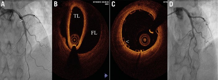
Figure 1. Angiographic and OCT images of SCAD before and after PCI with BVS. A) Coronary angiogram in AP cranial view showing a linear intraluminal filling defect (>) extending from LMCA to mid LAD. B) OCT image at LAD level showing total degloving of the intima and media from the adventitia. C) OCT at distal LMCA after BVS deployment showing well-apposed scaffold (<). D) Coronary angiogram in AP cranial view showing sealed flap. FL: false lumen; TL: true lumen.
Contrast staining in the LMCA prompted us to perform immediate wiring and OCT imaging in this life-threatening condition, which helped to identify the exact point of entry and the extent of the dissection, allowing appropriate deployment of the scaffold. As SCAD has a high tendency for spontaneous healing, we hypothesised that temporary scaffolding would facilitate this and avoid a permanent metallic implant1. A follow-up CT scan four months later showed a patent scaffold in the left main coronary artery with healed dissection.
Conflict of interest statement
The authors have no conflicts of interest to declare.
Supplementy data
Moving image 1. Initial angiogram showing dynamic LMCA compression and persistent contrast staining.
Moving image 2. Angiogram after wiring showing the dissection flap.
Moving image 3. Final angiogram after BVS and sealing of the flap.