IMAGE IN CARDIOLOGY

DOI: 10.4244/EIJV10I11A225

The “plastic healing concept”: implantation of bioabsorbable scaffolds in spontaneous coronary artery dissection

Ahmed Al Mamary, MD; Gilberto Dariol, MD; Massimo Napodano*, MD

A 45-year-old woman presented to our department because of inferior STEMI. A coronary angiogram showed patent coronary arteries with a near-occlusive smooth lesion from the mid to distal large obtuse marginal (OM), but otherwise normal vessels (Figure 1A). IVUS indicated the presence of intramural haematoma, with thrombosis of false lumen compressing the true lumen (Figure 1B, Moving image 1). Two bioabsorbable scaffolds (Absorb 3.5×12 mm+3.0×18 mm; Abbott Vascular, Santa Clara, CA, USA) were directly deployed with short overlap. Angiogram/IVUS revealed complete scaffold deployment, with fully apposed struts to the intimomedial membrane and no residual haematoma outside the scaffold area (Figure 1C, Figure 1D, Moving image 2). Six months later, a coronary computed tomography angiogram revealed the patency of the BVS with no residual haematoma of the vessel wall outside the scaffold area (Figure 1E, Figure 1F).

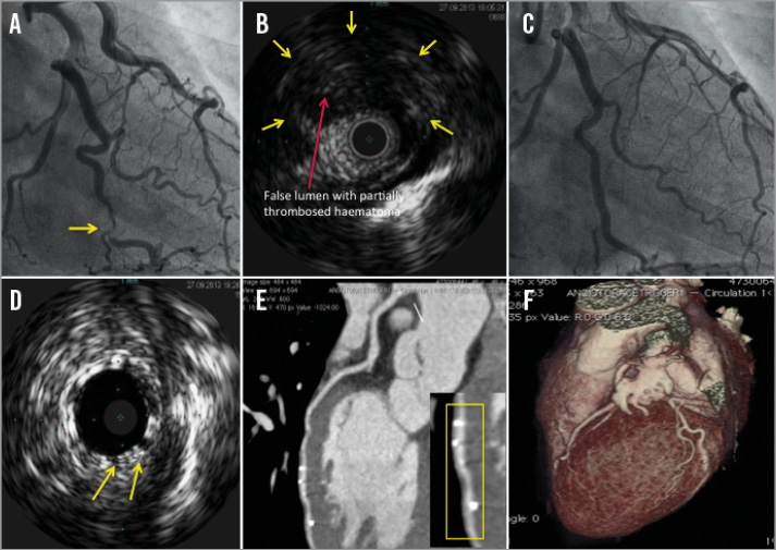
Figure 1. Imaging of SCAD before and after BVS implantation. A) Angiogram showing smooth lesion of the obtuse marginal (OM) (yellow arrowhead). B) IVUS shows false lumen (yellow arrowheads), with partially thrombosed haematoma, compressing the true lumen (red arrowhead). C) & D) Angiogram/IVUS after two bioabsorbable vascular scaffolds (BVS) had been implanted, showing full expansion of the scaffolds apposed to the intimomedial membrane. E) & F) Six-month computed tomography angiogram reveals the patency of the BVS (yellow rectangle), with no residual haematoma of the vessel wall outside the scaffold area.

Conflict of interest statement

The authors have no conflicts of interest to declare.

Online data supplement

Moving image 1. Intravascular ultrasound scan shows the presence of false lumen (9 o’clock to 2 o’clock) of the obtuse marginal branch with partially thrombosed haematoma, compressing the true lumen, without clear imaging of intimal tear appreciable.

Moving image 2. Intravascular ultrasound of the obtuse marginal branch after the implantation of two bioabsorbable vascular scaffolds, showing uniform full expansion of scaffolds apposed to the intima, with healing of abluminal haematoma.

Volume 10 Number 11
Mar 20, 2015
Volume 10 Number 11
View full issue


Key metrics

Suggested by Cory

INTERVENTIONAL FLASHLIGHT

10.4244/EIJ-D-17-00192 Oct 20, 2017
Long-term outcome of a spontaneous coronary artery dissection treated with a bioresorbable scaffold
free

SHORT REPORT

10.4244/EIJ-D-18-00519 Jan 18, 2019
Long-term follow-up of spontaneous coronary artery dissection treated with bioresorbable scaffolds
Macaya F et al
free

INTERVENTIONAL FLASHLIGHT

10.4244/EIJ-D-18-00035 Oct 12, 2018
Hybrid percutaneous treatment of iatrogenic coronary artery dissection complicating a spontaneous coronary artery dissection
García-Guimaraes M et al
free

IMAGE IN CARDIOLOGY

10.4244/EIJV11I1A14 May 19, 2015
Very late bioresorbable vascular scaffold thrombosis: a new clinical entity
Azzalini L et al
free
Trending articles
202.75

State-of-the-Art

10.4244/EIJ-D-21-00089 Jun 11, 2021
Intracoronary optical coherence tomography: state of the art and future directions
Ali ZA et al
free
87.2

State-of-the-Art

10.4244/EIJ-D-25-00266 Jan 19, 2026
Lesion stratification with intracoronary imaging
McGarvey M et al
free
47.45

NEW INNOVATION

10.4244/EIJ-D-15-00467 Feb 20, 2018
Design and principle of operation of the HeartMate PHP (percutaneous heart pump)
Van Mieghem NM et al
free
34.8

Original Research

10.4244/EIJ-D-25-01006 Mar 16, 2026
Clinical outcomes and haemodynamic response after blinded stress assessment of moderate aortic stenosis
Eerdekens R et al
24.15

Original Research

10.4244/EIJ-D-25-01370 May 21, 2026
Prognostic value of early haemodynamic valve deterioration after TAVI
Trimaille A et al
24.15

Original Research

10.4244/EIJ-D-25-01370 Jun 1, 2026
Prognostic value of early haemodynamic valve deterioration after TAVI
Trimaille A et al
23.2

State-of-the-Art

10.4244/EIJ-D-25-00874 Jun 1, 2026
TAVI and coronary interventions: indications, technical considerations, and clinical scenarios
Aquino Bruno H et al
free
22.2

Viewpoint

10.4244/EIJ-D-25-01066 May 4, 2026
Intracoronary imaging guidance for de novo coronary lesion treatment with drug-coated balloons
Amabile N et al
free
X

PCR
Impact factor: 9.5
2024 Journal Citation Reports®
Science Edition (Clarivate Analytics, 2025)
Online ISSN 1969-6213 - Print ISSN 1774-024X
© 2005-2026 Europa Group - All rights reserved