DOI: 10.4244/EIJV7I2A47

Healing of a coronary artery dissection detected by intravascular ultrasound and optical coherence tomography

Roberto Diletti, MD; Hector M. Garcia-Garcia, MD, PhD; Salvatore Brugaletta, MD; Josep Gomez Lara, MD; Patrick W. Serruys*, MD, PhD, FESC; Evelyn Regar, MD, PhD

A 56-year-old woman, active smoker with no other cardiovascular risk factors was admitted to hospital to undergo elective angioplasty on the proximal circumflex artery. A bioresorbable scaffold BVS 3.0×18mm was implanted. Post-implantation IVUS showed a residual coronary dissection at the distal edge of the scaffold (online Figure). In addition, OCT was performed showing an intimal flap reaching the first frame proximal to the take-off of the side branch distal to the scaffold (Figure1). The dissection was left untreated due to the lack of angiographic evidence of intimal flap and TIMI flow grade 3.1
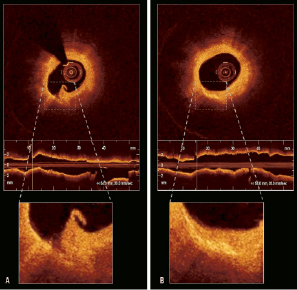
Figure 1. OCT performed after the procedure revealed a coronary dissection (Panel A). At six months follow-up, there was no evidence of dissection but an area of high reflectivity exactly in the same region according to the position of the side branch used as landmark (Panel B).

After six months, coronary angiogram, IVUS-VH and OCT were repeated showing a complete healing of the dissection, however by OCT there was a clearly defined region of high reflectivity (Figure1). We hypothesise that this increase in reflectivity could be related to a local increase in fibrotic tissue and inflammatory cells2 as a result of the wound healing process and should be considered as an intimal scar. There have been previous reports in the IVUS era describing the fate of edge dissections3. However, this is one of the first observations using OCT. The high sensitivity of this technology allows us to observe the mechanisms of repair of the dissection and could be used in longitudinal studies to observe the evolution over time of residual non-obstructive dissections. The study of these complications may have important clinical implications, in order to define which of these dissections deserve further treatment during the index procedure according to the follow-up findings.

Conflict of interest statement

The authors have no conflicts of interest to declare.

Online data supplement

Figure. After the procedure IVUS clearly identified a dissection at the distal edge of the scaffold but no evidence of dissection could be found at the angiogram (middle panels). At six month follow-up there was no angiographic and no IVUS evidence of dissection and virtual histology did not show any remarkable change in plaque composition (right panels).

Moving image 1. Oct run portraying the dissection post procedure.

Moving image 2. Oct run at six months with no evidence of dissection.
Volume 7 Number 2
Jun 30, 2011
Volume 7 Number 2
View full issue


Key metrics

On the same subject

Editorial

10.4244/EIJ-E-24-00010 Apr 15, 2024
Timing of revascularisation in acute coronary syndromes with multivessel disease – two sides of the same coin
Stähli B and Stehli J
free

Editorial

10.4244/EIJ-E-24-00006 Apr 15, 2024
The miracle of left ventricular recovery after transcatheter aortic valve implantation
Dauerman H and Lahoud R
free

Research Correspondence

10.4244/EIJ-D-23-01046 Apr 15, 2024
Feasibility and safety of transcaval venoarterial extracorporeal membrane oxygenation in severe cardiogenic shock
Giustino G et al

State-of-the-Art

10.4244/EIJ-D-23-00836 Apr 15, 2024
Renal denervation in the management of hypertension
Lauder L et al
free

Original Research

10.4244/EIJ-D-23-00643 Apr 15, 2024
A study of ChatGPT in facilitating Heart Team decisions on severe aortic stenosis
Salihu A et al
Trending articles
338.63

State-of-the-Art Review

10.4244/EIJ-D-21-00904 Apr 1, 2022
Antiplatelet therapy after percutaneous coronary intervention
Angiolillo D et al
free
295.45

Expert consensus

10.4244/EIJ-D-21-00898 Sep 20, 2022
Intravascular ultrasound guidance for lower extremity arterial and venous interventions
Secemsky E et al
free
226.03

State-of-the-Art Review

10.4244/EIJ-D-21-00426 Dec 3, 2021
Myocardial infarction with non-obstructive coronary artery disease
Lindahl B et al
free
209.5

State-of-the-Art Review

10.4244/EIJ-D-21-01034 Jun 3, 2022
Management of in-stent restenosis
Alfonso F et al
free
168.4

Expert review

10.4244/EIJ-D-21-00690 May 15, 2022
Crush techniques for percutaneous coronary intervention of bifurcation lesions
Moroni F et al
free
149.53

State-of-the-Art

10.4244/EIJ-D-22-00776 Apr 3, 2023
Computed tomographic angiography in coronary artery disease
Serruys PW et al
free
103.48

Expert consensus

10.4244/EIJ-E-22-00018 Dec 4, 2023
Definitions and Standardized Endpoints for Treatment of Coronary Bifurcations
Lunardi M et al
free
X

The Official Journal of EuroPCR and the European Association of Percutaneous Cardiovascular Interventions (EAPCI)

EuroPCR EAPCI
PCR ESC
Impact factor: 6.2
2022 Journal Citation Reports®
Science Edition (Clarivate Analytics, 2023)
Online ISSN 1969-6213 - Print ISSN 1774-024X
© 2005-2024 Europa Group - All rights reserved