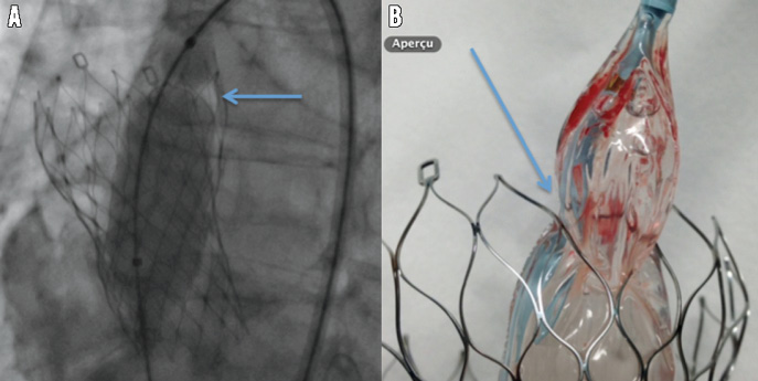
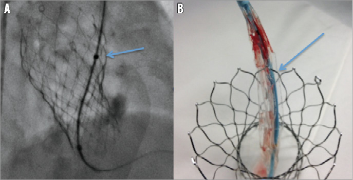
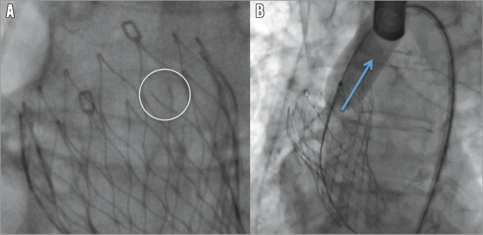
The occurrence of aortic regurgitation (AR) after transcatheter aortic valve implantation (TAVI) is common, and the consequences justify correcting grade 2 or more AR. During a standard TAVI procedure for an 85-year-old woman using a 26 mm self-expanding CoreValve® (Medtronic, Minneapolis, MN, USA) through a left femoral approach, a severe aortic regurgitation needed an additional balloon valvuloplasty. We used an AL2 Amplatz catheter (Terumo Corp., Tokyo, Japan), advanced through the prosthesis switched with a Super Stiff Amplatz (Boston Scientific, Marlborough, MA, USA). The initial balloon crossed the valve and was very gently inflated. After complete deflation, it was impossible to pull back the balloon (Moving image 1). The J guidewire had crossed a frame cell and was trapped in the upper angle of the nitinol frame cell. Ex vivo didactic pictures demonstrate the key issue (Online Figure 1). A new progressive reinflation broke the cell, dislodging the balloon (Figure 1, Online Figure 2, Moving image 2). The end-procedure angiogram confirmed a mild periprosthetic regurgitation. The immediate outcome was event-free. According to this experience, it seems to be safer to keep the guidewire inside the left ventricle during removal of the AccuTrak™ (Medtronic) and to pull it back definitively only after being certain there is no need for an additional dilatation.

Figure 1. Balloon reinflation to break the cell frame. A) The balloon is slowly inflated with the waist well identified inside the strut (blue arrow). B) Ex vivo illustration.
Conflict of interest statement
The authors have no conflicts of interest to declare.
Supplementary data
Moving image 1. The balloon is blocked through the cell frame.
Moving image 2. Dislodging the balloon.
` 
Online Figure 1. Balloon through the cell frame. A) The balloon has gone the wrong way through the strut of the cell frame (blue arrow). B) Ex vivo illustration.

Online Figure 2. The broken cell frame. A) The cell frame is broken (white circle). B) The balloon comes up (blue arrow).
Supplementary data
To read the full content of this article, please download the PDF.
Moving image 1. The balloon is blocked through the cell frame.
Moving image 2. Dislodging the balloon.

