The presence of a redundant Eustachian valve (EV) might be difficult to distinguish from a double atrial septum (DAS)1. The EV is generally anterior, whereas a DAS tends to be located posterior and to the left of the inferior vena cava2. We describe the percutaneous closure of an atrial septal defect (ASD) in a patient in whom intraprocedural transoesophageal echocardiography (TEE) showed a possible DAS not detected on previous TEE. Since the operators were not sure if the image corresponded to a redundant EV or a DAS, a pigtail catheter was advanced into the right ventricle (RV). The pigtail deflected the prominent tissue, confirming the diagnosis of a redundant EV (Figure 1). During the ASD occlusion, the pigtail was left inside the RV to avoid any tissue interference with the ASD occluder (Appendix Figure 1). The utility of a pigtail catheter to deflect the EV during percutaneous ASD occlusion has been reported previously. However, our case demonstrates the usefulness of an RV catheter not only to avoid tissue interference during ASD occlusion but also to distinguish between anatomical variations (Figure 1).
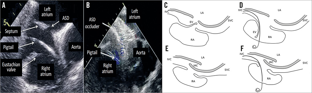
Figure 1. Differences between double atrial septum and redundant Eustachian valve. TEE image of the EV deflection with a pigtail (A) precluding any tissue interference with the ASD occluder (B). The insertion of a catheter into the RV changes the septum appearance in a redundant EV (C&D) but not in a DAS (E&F). EV: Eustachian valve; IAC: interatrial chamber; IVC: inferior vena cava; LA: left atrium; RA: right atrium; SVC: superior vena cava.
Conflict of interest statement
The authors have no conflicts of interest to declare.
Supplementary data
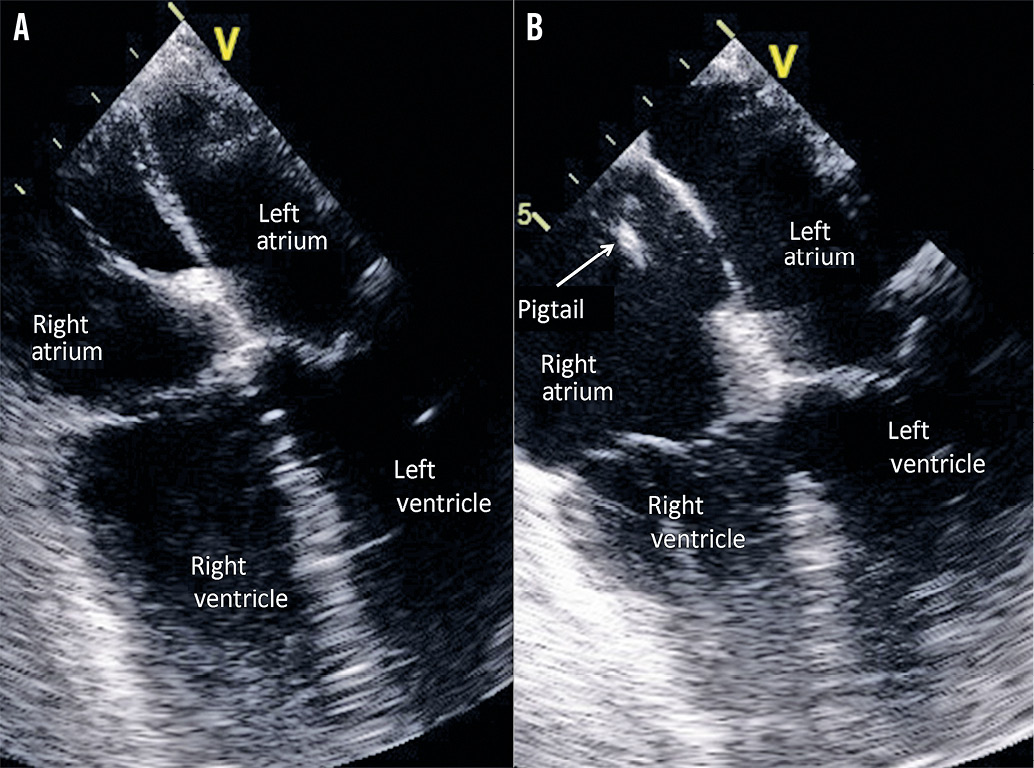
Appendix Figure 1. TEE image. The possible DAS (A) was ruled out after deflecting a redundant EV with a pigtail catheter (B).