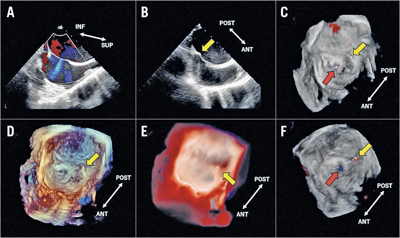
Figure 1. 2D- and 3D-TEE images of the atrial septum during the MitraClip procedure. The red and yellow arrows indicate the previous iASD and tenting point (new iASD), respectively.
A 60-year-old man who had catheter ablation due to atrial fibrillation underwent transcatheter mitral valve repair due to severe secondary mitral regurgitation. The procedure was performed under general anaesthesia with two-dimensional (2D) and three-dimensional (3D) transoesophageal echocardiography (TEE) using the EPIQ CVx/X8-2t ultrasound system (Philips Medical Systems, Andover, MA, USA). During the procedure, 2D-TEE demonstrated the previous iatrogenic atrial septal defect (iASD) at the mid-anterior location of the fossa ovalis (Figure 1A). To obtain sufficient height from the mitral annulus, the tip of a radiofrequency transseptal needle was positioned at the mid-posterior location of the fossa ovalis (Figure 1B for the 2D-TEE, Figure 1C, Moving image 1 for the 3D-TEE colour image). Although the 3D-TEE colour and classic 3D-TEE (Figure 1D, Moving image 2) images are able to visualise the tenting point, the 3D-TrueVue (Philips Medical Systems) image, which is a new technology that produces virtual light, enabled clarifying the tenting point on the left atrial side by drawing shadows (Figure 1E, Moving image 3). There was sufficient distance (13.6 mm; Supplementary Figure 1) between the iASD and the tenting point on the 3D-TrueVue image, which was measured using the combination of the 3D-TrueVue image and the 3D-TEE colour image, and thus puncture was performed. Subsequently, two MitraClip® devices (Abbott Vascular, Santa Clara, CA, USA) were successfully implanted. After removal of the system, the two iASDs (old and new) with left-to-right shunts were confirmed using the 3D-TEE colour image (Figure 1F, Moving image 4).
The puncture point should be at a sufficient distance from the iASD to avoid a merged large iASD that may impact on the stability of the guide catheter of the MitraClip system. In addition, significant iASD after using the MitraClip is associated with worse outcomes1. TrueVue is a new 3D realistic rendering image that provides shadowing from the virtual lighting that can improve depth perception within a shallower depth in the 3D images2. This new technology may allow a non-echocardiography specialist to visualise the tenting location more clearly than with the classic 3D-TEE imaging, thereby resulting in better communication within the Heart Team during the MitraClip procedure. This is useful in patients with a history of catheter ablation or abnormal anatomy of the atrial septum.
Conflict of interest statement
The authors have no conflicts of interest to declare.
Supplementary data
To read the full content of this article, please download the PDF.
Moving image 1. 3D-TEE colour moving image.
Moving image 2. Classic 3D-TEE moving image.
Moving image 3. 3D-TEE TrueVue moving image.
Moving image 4. 3D-TEE colour moving image after removal of the MitraClip system.