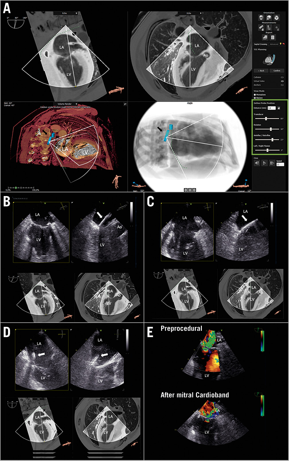
Figure 1. CT-derived transoesophageal echocardiographic views for direct transcatheter mitral valve annuloplasty. A) The virtual TEE probe (black arrow) placed in the oesophagus and gastric fundus on the sagittal MDCT view and predicted fluoroscopy view. On the right side are shown the settings (green box) that can be adjusted to derive TEE views from MDCT data. The ideal MDCT-derived TEE views can be obtained and reproduced during the procedure to guide the catheter (white arrow) and place the anchors in the predicted position (B-D). E) The reduction of mitral regurgitation grade from severe (upper image) to moderate (lower image) after successful transcatheter mitral valve annuloplasty with the Cardioband system, size D. Ao: aorta; LA: left atrium; LV: left ventricle
Multidetector row computer tomography (MDCT) and transoesophageal echocardiography (TEE) are the imaging modalities of choice for patient selection and planning of transcatheter valvular interventions1. For transcatheter mitral valve annuloplasty with the Cardioband™ system (Edwards Lifesciences, Irvine, CA, USA), high spatial resolution data from MDCT are required for accurate measurement of the mitral valve annulus (MVA) to select the device size and for assessment of the distance between MVA and circumflex artery to ensure safe implantation of the anchors of the device1. TEE is key to assess the mechanism and severity of mitral regurgitation. However, TEE has lower spatial resolution compared with MDCT and, in some patients, it might be difficult to image the location where the anchors of the Cardioband system should be placed. A dedicated software (3mensio; Pie Medical Imaging BV, Maastricht, the Netherlands) for post-processing MDCT data allows reproducing TEE views which may be compared to the TEE images needed for procedural guidance (Figure 1A-Figure 1D). This novel feature can be used for teaching purposes and may provide several advantages in the imaging key steps to guide transcatheter mitral valve annuloplasty. Specifically, it may help to identify the best TEE views for transseptal puncture and to image the predefined MDCT anchoring sites of the device, potentially allowing faster and better alignment of the anchors during the procedure. The virtual TEE probe (Figure 1A, black arrow) can be placed in the oesophagus and its angulation and anteflexion or retroflexion can be adjusted to obtain the desired MDCT-derived TEE view (green box). The software also allows visualisation of the height of the probe on the predicted fluoroscopic views that could be challenging to reproduce during the procedure. We present the case of a 66-year-old patient with ischaemic severe mitral regurgitation and New York Heart Association (NYHA) Class III heart failure symptoms who underwent transcatheter mitral valve annuloplasty due to high operative risk. In the preoperative TEE evaluation, the imaging of the posterolateral MVA was particularly challenging. The MDCT-derived TEE views helped to identify the best TEE images to guide the catheter (Figure 1B-Figure 1D, white arrow) during the procedure. The procedure was successful with a significant reduction in the annular diameters and mitral regurgitation (Figure 1E), accompanied by an improvement in heart failure symptoms (NYHA Class I).
Funding
The Department of Cardiology of the Leiden University Medical Center received research grants from Abbott Vascular, Bayer, BioVentrix, Medtronic, Biotronik, Boston Scientific, GE Healthcare and Edwards Lifesciences.
Conflict of interest statement
N. Ajmone Marsan reports speaker fees from Abbott Vascular. V. Delgado reports personal fees from Abbott Vascular, MSD, Novartis, Edwards Lifesciences, Medtronic, and GE Healthcare. The other authors have no conflicts of interest to declare.
Supplementary data
To read the full content of this article, please download the PDF.