A 70-year-old woman with known complete thoracic paraplegia, chronic obstructive pulmonary disease and gastrointestinal bleedings was referred for a MitraClip® (Abbott Vascular, Santa Clara, CA, USA) procedure for symptomatic mitral regurgitation (MR). Echocardiography showed moderate-severe MR due to prolapse of degenerated leaflets (Barlow’s disease) (Online Figure 1, Online Figure 2, Moving image 1, Moving image 2).
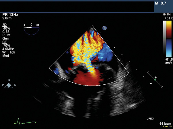
The first clip was implanted on the origin of the largest MR jet in a lateral position, reducing MR to moderate with a mean gradient of 3-4 mmHg. During closure of the second clip, complete sealing of the mitral valve (MV) occurred with concomitant loss of blood pressure. Echocardiography showed spontaneous contrast in the left atrium (LA) and complete collapse of the left ventricle (LV) (Figure 1, Moving image 3). After re-opening the clip, recovery of haemodynamic state occurred.
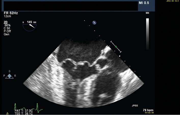
Figure 1. Mid-oesophageal transoesophageal long-axis view after closure of the second MitraClip showing dense spontaneous contrast in the LA and a complete collapse of the LV.
This case emphasises that patients with Barlow’s disease may not be suitable for the MitraClip. The highly mobile MV frequently results in residual MR, while grasping the redundant tissue increases the risk of relevant MV stenosis.
Conflict of interest statement
M. Swaans and J. Van der Heyden are proctors for Abbott. The other authors have no conflicts of interest to declare.
Online data supplement
Moving image 3. Mid-oesopheageal transoesophageal long-axis view after closure of the second MitraClip showing dense spontaneous contrast in the LA and a complete collapse of the LV.
Online Figure 1. Mid-oesophageal transoesophageal four-chamber view showing prolapse of both leaflets.
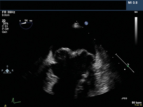
Online Figure 2. Mid-oesophageal transoesophageal four-chamber view with colour showing prolapse of both leaflets causing moderate-severe MR.
Supplementary data
To read the full content of this article, please download the PDF.
Moving image 1. Mid-oesophageal transoesophageal four-chamber view with and without colour showing prolapse of both leaflets causing moderate-severe MR.
Moving image 2. Mid-oesophageal transoesophageal four-chamber view with and without colour showing prolapse of both leaflets causing moderate-severe MR.
Moving image 3. Mid-oesopheageal transoesophageal long-axis view after closure of the second MitraClip showing dense spontaneous contrast in the LA and a complete collapse of the LV.