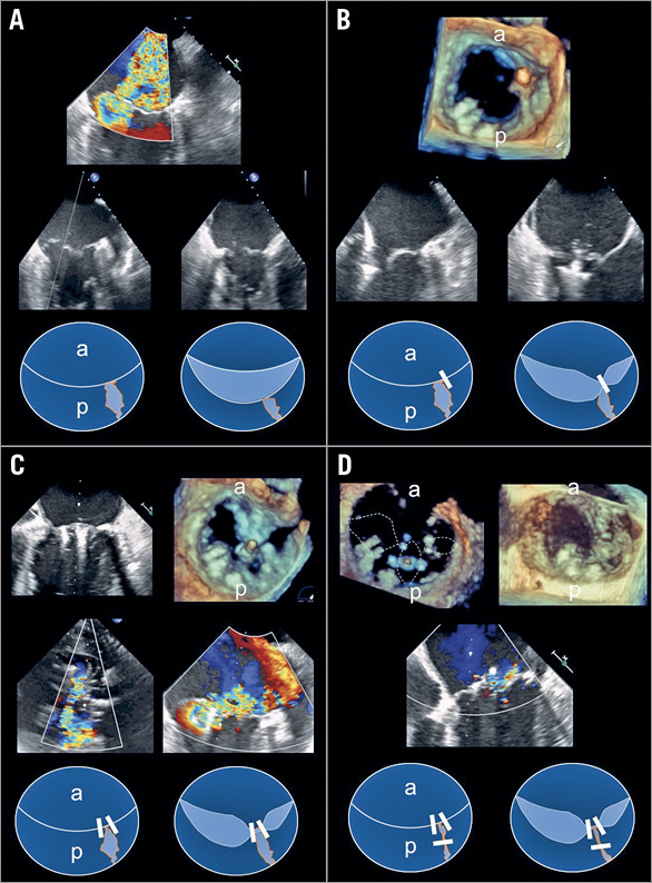

Figure 1. Echocardiographic and schematic overview of the major steps in the procedure. A) Colour Doppler showing severe mitral regurgitation (MR), B-mode image of intercommissural and grasping view displaying the underlying pathology, schematic en face view of the mitral valve. B) 3D en face view with first clip in place, clip orientation in intercommissural and grasping view before grasping, schematic view after first clip. C) Intercommissural and en face view with second clip in place, residual MR, schematic view after second clip. D) En face view showing clip orientation within the cleft before and after grasping, residual minor MR after third clip, schematic view after third clip.
A 60-year-old male was referred to our intensive care unit with progressive respiratory failure and pulmonary haemorrhage. Transoesophageal echocardiography revealed severe degenerative mitral regurgitation (MR) due to a flail of the P3/P2 segment complicated by a cleft of the posterior leaflet reaching to the base of the posterior mitral leaflet (PML) (Figure 1A , Moving image 1-Moving image 2B). An interventional approach using the MitraClip® system (Abbott Vascular, Santa Clara, CA, USA) was chosen by the Heart Team since the patient was unsuitable for a surgical repair due to significant comorbidities (EuroSCORE II 17.6%). The first clip was positioned medial (Figure 1B), the second clip lateral to the cleft in order to address the flailing posterior leaflet and stabilise the cleft (Figure 1C). However, significant residual MR remained due to the persistent regurgitation within the cleft. Therefore, a third clip was placed orthogonally to the edges of the cleft within the PML (Moving image 3), resulting in minor residual MR with a mean transmitral gradient of 3 mmHg (Figure 1D, Moving image 4-Moving image 6). One year after the procedure the patient is able to go hiking without experiencing dyspnoea. Transthoracic echocardiography continues to show a persistent functional repair with only mild MR.
Several groups have described a technique using two convergent clips on both sides of the cleft1-3. Indeed, we used this technique as the initial approach in our case and went on to address the cleft itself directly only after an unsatisfactory result. To our knowledge, our case is the first report of an intra-leaflet clipping procedure.
Conflict of interest statement
T. Wengenmayer has received proctoring fees and research support from Abbott Vascular. The other authors have no conflicts of interest to declare.
Supplementary data
To read the full content of this article, please download the PDF.
Moving image 1. Mitral regurgitation before the procedure.
Moving image 2A. Anatomy of the cleft between segments 2 and 3 of the posterior mitral leaflets.
Moving image 2B. 2D mitral valve anatomy before intervention.
Moving image 3. MitraClip orientation towards the cleft.
Moving image 4. Anatomy of the mitral valve at the end of the procedure.
Moving image 5. Mitral regurgitation after the procedure.
Moving image 6. Location of the implanted clips.

