IMAGE IN CARDIOLOGY

DOI: 10.4244/EIJV12I10A212

Triple orifice as a novel strategy in interventional reconstruction of a mitral pseudo cleft

Tobias Wengenmayer*, MD; Qian Zhou, MD; Christoph Bode, MD; Manfred Zehender, MD

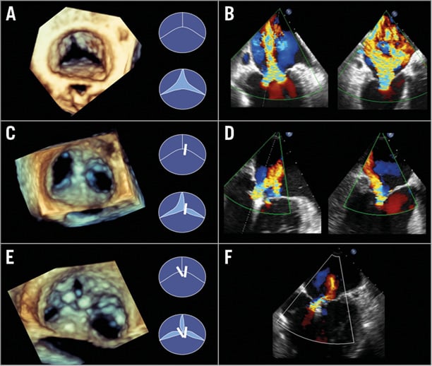
An 84-year-old male with a history of ischaemic cardiomyopathy was admitted with advanced NYHA Class IV symptoms. Transoesophageal echocardiography revealed a severe functional mitral regurgitation (MR) with a pseudo cleft in the P2 segment (Panel A, Panel B). Surgical repair was not possible due to his comorbidities (EuroSCORE 54.41%), thus an interventional approach using the MitraClip® system (Abbott Vascular, Santa Clara, CA, USA) was chosen by the Heart Team.

A clip was positioned in the A2/P2 region perpendicular to the commissure and lateral to the cleft, resulting in stabilisation of the cleft and reducing MR to moderate (Panel C, Panel D). The patient was discharged three days after the procedure with NYHA Class II symptoms.

Eighteen weeks later, the patient was re-admitted with severe dyspnoea. Echocardiography revealed moderate to severe MR (Panel D). Placing a second clip parallel to the first clip did lead to a significant reduction in mitral regurgitation but also caused an excessive rise of transmitral gradient. Therefore, we placed the second clip with a 45-degree angle to the first clip in order to create a triple orifice (Panel E). MR was reduced from moderate to severe to mild with a mean transmitral gradient of 5 mmHg (Panel F). Two days after the procedure, our patient was discharged with NYHA functional Class I.

To our knowledge, this is the first report of a MitraClip-induced triple orifice in a case of mitral pseudo cleft. By using the cleft itself as an orifice, mitral regurgitation could be effectively reduced without affecting the mean transmitral gradient (Moving image 1-Moving image 4).

Conflict of interest statement

The authors have no conflicts of interest to declare.

Supplementary data

Moving image 1. Atrial view prior to intervention (pseudo cleft in P2).

Moving image 2. Ventricular view prior to intervention (pseudo cleft in P2).

Moving image 3. Atrial view after 1st clip.

Moving image 4. Atrial view after 2nd clip.

Supplementary data

To read the full content of this article, please download the PDF.

Atrial view prior to intervention (pseudo cleft in P2).

Ventricular view prior to intervention (pseudo cleft in P2).

Atrial view after 1 clip.

Atrial view after 2 clip.
Volume 12 Number 10
Nov 20, 2016
Volume 12 Number 10
View full issue


Key metrics

Suggested by Cory

Image – Interventional flashlight

10.4244/EIJ-D-18-00772 Mar 20, 2019
Clipping within the leaflet: a novel approach to complex mitral regurgitation pathologies
Wengenmayer T et al
free

IMAGE IN CARDIOLOGY

10.4244/EIJ-D-16-00395 Feb 3, 2017
A2-P3 oblique clipping for the treatment of severe mitral regurgitation in the presence of mitral valve cleft and flail
Tarantini G et al
free

IMAGE IN CARDIOLOGY

10.4244/EIJY14M07_05 Jul 20, 2015
MitraClip repair of a “trileaflet” regurgitant mitral valve
Freixa X et al
free

IMAGE IN CARDIOLOGY

10.4244/EIJV12I8A173 Oct 10, 2016
Challenging mitral clefts with MitraClip: the convergent clips strategy
Taramasso M and Maisano F
free

MITRAL VALVE INTERVENTIONS

10.4244/EIJV12SYA14 Sep 18, 2016
The MitraClip system: strategies for optimal patient selection and optimised results
Grasso C and Ince H
free

INTERVENTIONAL FLASHLIGHT

10.4244/EIJ-D-16-00943 Oct 20, 2017
Transcatheter mitral annuloplasty to treat residual mitral regurgitation after MitraClip implantation
Brüstle K et al
free

10.4244/EIJV11SWA11 Sep 17, 2015
The MitraClip transcatheter mitral valve repair system
Grasso C and Baldus S
free
Trending articles
151.43

State-of-the-Art

10.4244/EIJ-D-22-00776 Apr 3, 2023
Computed tomographic angiography in coronary artery disease
Serruys PW et al
free
55.9

Clinical research

10.4244/EIJ-D-22-00621 Feb 20, 2023
Long-term changes in coronary physiology after aortic valve replacement
Sabbah M et al
free
54.9

Expert review

10.4244/EIJ-D-21-01010 Jun 24, 2022
Device-related thrombus following left atrial appendage occlusion
Simard T et al
free
43.75

Clinical Research

10.4244/EIJ-D-21-01091 Aug 5, 2022
Lifetime management of patients with symptomatic severe aortic stenosis: a computed tomography simulation study
Medranda G et al
free
39.95

Clinical research

10.4244/EIJ-D-22-00558 Feb 6, 2023
Permanent pacemaker implantation and left bundle branch block with self-expanding valves – a SCOPE 2 subanalysis
Pellegrini C et al
free
X

The Official Journal of EuroPCR and the European Association of Percutaneous Cardiovascular Interventions (EAPCI)

EuroPCR EAPCI
PCR ESC
Impact factor: 7.6
2023 Journal Citation Reports®
Science Edition (Clarivate Analytics, 2024)
Online ISSN 1969-6213 - Print ISSN 1774-024X
© 2005-2024 Europa Group - All rights reserved