IMAGE IN CARDIOLOGY

DOI: 10.4244/EIJ-D-16-00395

A2-P3 oblique clipping for the treatment of severe mitral regurgitation in the presence of mitral valve cleft and flail

Giuseppe Tarantini*, MD, PhD; Giulia Masiero, MD; Umberto Cucchini, MD, PhD; Luca Nai Fovino, MD

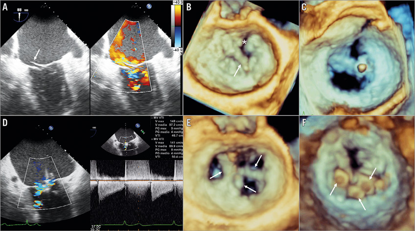
An 86-year-old male was admitted to our institution for dyspnoea. Three-dimensional transoesophageal echocardiography (TEE) showed severe mitral regurgitation (MR) caused by both A2 prolapse with a flail due to cord rupture and a cleft in the anterior mitral leaflet (Panel A, Panel B, Moving image 1). Given the prohibitive surgical risk, the patient was considered for MitraClip® (Abbott Vascular, Santa Clara, CA, USA) implantation.

After several unsuccessful attempts to perform an initial A2-P2 central grasp, the clip was rotated counterclockwise grasping the P3 scallop and the point of connection of a ruptured primary cord to the A2/A1 region, bridging the proximal part of the anterior cleft (Panel C, Moving image 2). Leaflet insertion assessment utilising intercommissural and left ventricular outflow tract views ensured that grasping was sufficient to prevent clip detachment. Following the implantation of two other clips (grasping A2/P2 and A1/P1), MR was reduced to grade 1+, with no significant residual mitral stenosis (Panel D-Panel F, Moving image 3, Moving image 4). Intraoperative TEE monitoring was performed with 3D atrial and X-plane from intercommissural views. Three-month follow-up showed persistence of good echocardiographic (MR grade 1+) and clinical (NYHA Class I) results.

This is the first report of MitraClip implantation in a rare and unfavourable anatomical condition, such as the coexistence of cleft and flail in the anterior mitral leaflet. The presence of a cleft is usually regarded as a contraindication to percutaneous repair, since the clip could worsen MR by displacing the clefted leaflet. Our “oblique” clipping allowed easier implantation of two other clips and the achievement of near complete MR reduction.

Acknowledgements

The authors would like to thank E. Adamovic (Engineer) for her precious technical support.

Conflict of interest statement

The authors have no conflicts of interest to declare.

Supplementary data

Moving image 1. Preprocedural 3D TEE atrial view of the mitral valve.

Moving image 2. Intraprocedural 3D TEE after the first “oblique” clipping.

Moving image 3. Postprocedural 3D TEE atrial view of the mitral valve.

Moving image 4. Postprocedural TEE Doppler showing near complete MR reduction.

Supplementary data

To read the full content of this article, please download the PDF.

video1
video2
video3
video4
Volume 12 Number 15
Feb 3, 2017
Volume 12 Number 15
View full issue


Key metrics

On the same subject

Image – Interventional flashlight

10.4244/EIJ-D-18-00772 Mar 20, 2019
Clipping within the leaflet: a novel approach to complex mitral regurgitation pathologies
Wengenmayer T et al
free

IMAGE IN CARDIOLOGY

10.4244/EIJV12I8A173 Oct 10, 2016
Challenging mitral clefts with MitraClip: the convergent clips strategy
Taramasso M and Maisano F
free

IMAGE IN CARDIOLOGY

10.4244/EIJV12I10A212 Nov 20, 2016
Triple orifice as a novel strategy in interventional reconstruction of a mitral pseudo cleft
Wengenmayer T et al
free

Expert review

10.4244/EIJ-D-20-00883 Nov 20, 2020
Percutaneous mitral valve leaflet repair: ongoing directions and future perspectives
Maisano F and Taramasso M
free

Image – Interventional flashlight

10.4244/EIJ-D-19-00897 Jul 17, 2020
Staged clasping with PASCAL device for mitral regurgitation with leaflet laceration
Sugiura A et al
free

MITRAL VALVE INTERVENTIONS

10.4244/EIJV12SYA14 Sep 18, 2016
The MitraClip system: strategies for optimal patient selection and optimised results
Grasso C and Ince H
free

Image – Interventional flashlight

10.4244/EIJ-D-23-00596 Nov 17, 2023
Electrosurgical laceration and stabilisation of three clip devices (ELASTA-Clip) to enable transcatheter mitral valve implantation
Curio J et al
Trending articles
281.53

State-of-the-Art Review

10.4244/EIJ-D-21-00695 Nov 19, 2021
Transcatheter treatment for tricuspid valve disease
Praz F et al
free
243.2

State of the art

10.4244/EIJ-D-21-01117 Sep 20, 2022
Recanalisation of coronary chronic total occlusions
Di Mario C et al
free
208.35

State-of-the-Art Review

10.4244/EIJ-D-21-01034 Jun 3, 2022
Management of in-stent restenosis
Alfonso F et al
free
168.7

Translational research

10.4244/EIJ-D-21-00824 May 15, 2022
Bench test and in vivo evaluation of longitudinal stent deformation during proximal optimisation
Toth GG et al
free
167.05

Expert review

10.4244/EIJ-D-21-00690 May 15, 2022
Crush techniques for percutaneous coronary intervention of bifurcation lesions
Moroni F et al
free
151.03

State-of-the-Art

10.4244/EIJ-D-22-00776 Apr 3, 2023
Computed tomographic angiography in coronary artery disease
Serruys PW et al
free
118

Translational research

10.4244/EIJ-D-22-00718 Jun 5, 2023
Preclinical evaluation of the degradation kinetics of third-generation resorbable magnesium scaffolds
Seguchi M et al
110.35

Viewpoint

10.4244/EIJ-E-22-00007 May 15, 2022
TAVI at 20: how a crazy idea led to a clinical revolution
Eltchaninoff H et al
free
X

The Official Journal of EuroPCR and the European Association of Percutaneous Cardiovascular Interventions (EAPCI)

EuroPCR EAPCI
PCR ESC
Impact factor: 6.2
2022 Journal Citation Reports®
Science Edition (Clarivate Analytics, 2023)
Online ISSN 1969-6213 - Print ISSN 1774-024X
© 2005-2024 Europa Group - All rights reserved