Image – Interventional flashlight

DOI: 10.4244/EIJ-D-18-00556

First experience with the MitraClip XTR device for extensive mitral valve prolapse (Barlow’s disease)

Karolina Weinmann, MD; Sinisa Markovic, MD; Wolfgang Rottbauer, MD; Mirjam Keßler*, MD

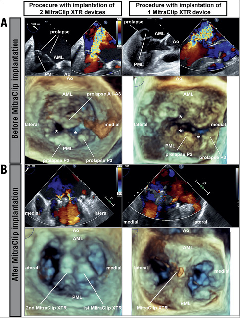
Figure 1. Transoesophageal echocardiography before and after MitraClip implantation. A) Before MitraClip implantation. B) After MitraClip implantation. * Structural defect. AML: anterior mitral leaflet; Ao: aortic valve; PML: posterior mitral leaflet

Various surgical strategies exist for Barlow’s disease (extensive mitral valve prolapse >1 segment, leaflet thickening)1. However, interventional therapy using the MitraClip® (Abbott Vascular, Santa Clara, CA, USA) in high-risk surgical candidates with Barlow’s disease is complex: either highly mobile leaflets lead to relevant residual mitral regurgitation (MR) or extensive grasping of redundant leaflet tissue might result in mitral stenosis2. In 2018 the MitraClip NTR and XTR (both Abbott Vascular) were launched, featuring technical modifications of the delivery catheter (shaft extension increased by 1.5 cm, more direct, predictable trajectory during positioning) and lock mechanism. In the MitraClip XTR, arm length has been increased from 9 mm to 12 mm and coaptation length and grasping is facilitated by two additional rows of frictional elements at the grippers. So far, the MitraClip XTR has been employed predominantly in functional MR and severely impaired left ventricular function3.

We used the MitraClip XTR in four patients with Barlow’s disease because of its increased coaptation length facilitating grasping of redundant tissue of prolapsing segments. All patients had severe MR (effective regurgitant orifice area of 0.6-2.2 cm2) with prolapse of >1 segment (6-13 mm above the annulus) (Figure 1A). The left atria were enlarged concomitant with severe MR, easing device positioning. Mitral annulus dilatation was not a prerequisite for the MitraClip XTR. By deployment of the MitraClip XTR at the main prolapse site, leaflet mobility was stabilised. Significant reduction to mild MR was achieved in one patient. In the other patients, a second MitraClip (NTR or XTR, depending on the length of the remaining leaflets) was placed at the centre of the remaining regurgitation to stabilise the leaflets further; this achieved sufficient MR reduction. No relevant transmitral gradients (mean pressure gradient 2-6 mmHg) occurred, even after implantation of a second MitraClip (Figure 1B).

Interventional treatment of Barlow’s disease is challenging due to the high mobility of the mitral leaflets; however, the latest MitraClip XTR renders the beneficial use of MitraClip devices possible.

Conflict of interest statement

The authors have no conflicts of interest to declare.

Volume 14 Number 12
Dec 20, 2018
Volume 14 Number 12
View full issue


Key metrics

On the same subject

Expert review

10.4244/EIJ-D-20-00883 Nov 20, 2020
Percutaneous mitral valve leaflet repair: ongoing directions and future perspectives
Maisano F and Taramasso M
free

10.4244/EIJV11SWA11 Sep 17, 2015
The MitraClip transcatheter mitral valve repair system
Grasso C and Baldus S
free

MITRAL VALVE INTERVENTIONS

10.4244/EIJV12SYA14 Sep 18, 2016
The MitraClip system: strategies for optimal patient selection and optimised results
Grasso C and Ince H
free

10.4244/EIJV11SWA10 Sep 17, 2015
Transcatheter mitral valve repair: a brief review
Barbanti M et al
free

EXPERT REVIEW

10.4244/EIJ-D-18-00511 Sep 7, 2018
Transcatheter mitral valve repair: review of the clinical evidence
Taramasso M et al
free

10.4244/EIJV11SWA14 Sep 17, 2015
Transcatheter direct mitral valve annuloplasty: a brief review
Feldman T and Guerrero M
free

EXPERT REVIEW

10.4244/EIJ-D-17-00505 Sep 24, 2017
Mitral valve: repair/clips/cinching/chordae
Vahanian A et al
free

Image – Interventional flashlight

10.4244/EIJ-D-23-00596 Nov 17, 2023
Electrosurgical laceration and stabilisation of three clip devices (ELASTA-Clip) to enable transcatheter mitral valve implantation
Curio J et al
Trending articles
281.53

State-of-the-Art Review

10.4244/EIJ-D-21-00695 Nov 19, 2021
Transcatheter treatment for tricuspid valve disease
Praz F et al
free
243.2

State of the art

10.4244/EIJ-D-21-01117 Sep 20, 2022
Recanalisation of coronary chronic total occlusions
Di Mario C et al
free
208.35

State-of-the-Art Review

10.4244/EIJ-D-21-01034 Jun 3, 2022
Management of in-stent restenosis
Alfonso F et al
free
168.7

Translational research

10.4244/EIJ-D-21-00824 May 15, 2022
Bench test and in vivo evaluation of longitudinal stent deformation during proximal optimisation
Toth GG et al
free
167.05

Expert review

10.4244/EIJ-D-21-00690 May 15, 2022
Crush techniques for percutaneous coronary intervention of bifurcation lesions
Moroni F et al
free
151.03

State-of-the-Art

10.4244/EIJ-D-22-00776 Apr 3, 2023
Computed tomographic angiography in coronary artery disease
Serruys PW et al
free
118

Translational research

10.4244/EIJ-D-22-00718 Jun 5, 2023
Preclinical evaluation of the degradation kinetics of third-generation resorbable magnesium scaffolds
Seguchi M et al
110.35

Viewpoint

10.4244/EIJ-E-22-00007 May 15, 2022
TAVI at 20: how a crazy idea led to a clinical revolution
Eltchaninoff H et al
free
X

The Official Journal of EuroPCR and the European Association of Percutaneous Cardiovascular Interventions (EAPCI)

EuroPCR EAPCI
PCR ESC
Impact factor: 6.2
2022 Journal Citation Reports®
Science Edition (Clarivate Analytics, 2023)
Online ISSN 1969-6213 - Print ISSN 1774-024X
© 2005-2024 Europa Group - All rights reserved