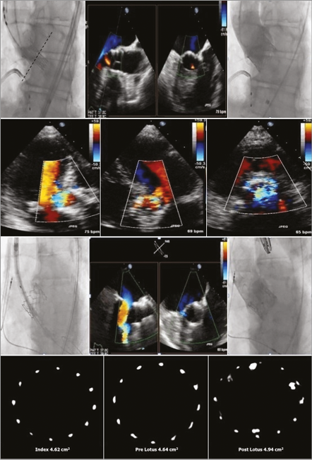
An 88-year-old male underwent a transcatheter aortic valve implantation with a 26 mm Edwards SAPIEN 3 (S3) valve (Edwards Lifesciences Inc., Irvine, CA, USA) with mild residual paravalvular regurgitation (PAR) after post-dilatation with a 26 mm balloon (Moving image 1). The upper row of Figure 1 illustrates the S3 positioning during expansion (left) with mild PAR by transoesophageal echocardiography (middle) and contrast angiography (right). Transthoracic echocardiography at follow-up (second row) revealed severe PAR (Moving image 2). The pathophysiology of this deterioration to severe PAR is uncertain. S3 sizing and positioning seemed adequate and recoil was excluded. Damage to the S3 integrity by post-dilatation cannot be ruled out. A transcatheter valve-in-S3 implantation with a 27 mm Lotus™ valve (Boston Scientific, Marlborough, MA, USA) followed. The third row displays the Lotus positioning within the S3 (left) and the final result (right) with trivial PAR (middle) (Moving image 3). The bottom row illustrates the mid portion of the frame derived from rotational angiography, confirming identical S3 area at the end of the index procedure and prior to Lotus implantation two months later (no recoil). Note the significant increase in area after Lotus implantation. The Lotus valve is mechanically deployed with high radial force and an adaptive seal. It is completely repositionable and retrievable even after full expansion. These intrinsic features may help optimise transcatheter valve-in-valve procedures.

Figure 1. Fluoroscopy and transoesophageal illustrations as explained in the text. Bottom row displays the cross-sectional view derived from the 3D reconstruction of the rotational angiography using dedicated Siemens software.
Guest Editor
This paper was guest edited by Alec Vahanian, MD; Cardiology Department, Bichat Hospital, Paris, France.
Conflict of interest statement
N. Van Mieghem has received research grants from Edwards Lifesciences, Medtronic and Boston Scientific. P. de Jaegere is a proctor for St. Jude Medical and Boston Scientific. The other authors have no conflicts of interest to declare. The Guest Editor is a consultant for Edwards Lifesciences.
Online data supplement
Moving image 1. Transoesophageal echocardiography after 26 mm S3 implantation shows mild PAR.
Moving image 2. Transthoracic echocardiography in parasternal short-axis view six days after TAVI shows severe PAR.
Moving image 3. Transoesophageal echocardiography after Lotus-in-S3 shows only trivial PAR.

