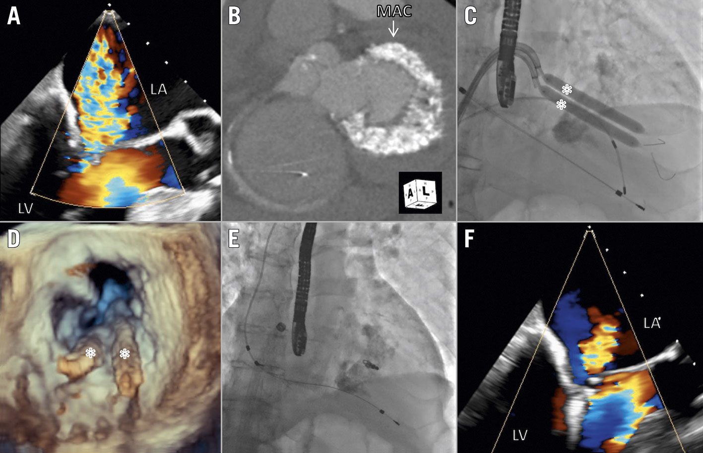
Figure 1. Procedural steps of lithotripsy-facilitated mitral edge-to-edge repair. A) Baseline echocardiogram demonstrating severe mitral regurgitation (MR). B) CT scan demonstrating severe mitral annular calcification. C) Fluoroscopy of lithotripsy balloons across the mitral valve. D) 3D echocardiogram of the lithotripsy balloons apposed to the calcified posterior mitral annulus and leaflet. E) Fluoroscopy after MitraClip. F) Echocardiogram demonstrating mild MR after the procedure. LA: left atrium; LV: left ventricle; MAC: mitral annular calcification; * lithotripsy balloon.
A 71-year-old man presented with persistent New York Heart Association (NYHA) Functional Class III dyspnoea after a recent heart failure hospitalisation. His past history included atrial fibrillation, hypertension and a permanent pacemaker. Echocardiography demonstrated severe calcific mitral regurgitation (MR) with valve area 2.2 cm2 and mean gradient 5 mmHg, with normal left ventricular (LV) function (Figure 1A, Moving image 1). The degree of mitral annular calcification (MAC) precluded surgery (Figure 1B), and his anatomy was unsuitable for transcatheter mitral valve replacement. After the Heart Team’s review, the patient underwent balloon lithotripsy-facilitated transcatheter edge-to-edge repair (TEER).
After the transseptal puncture, a MitraClip NT (Abbott) was initially placed at A2-P2, but was associated with moderate mitral regurgitation (MR) and mean gradient 6 mmHg, and thus was removed. The MitraClip guide catheter was exchanged for two Agilis steerable catheters (Abbott) in the left atrium. Subsequently, two 7 mm × 60 mm lithotripsy balloons (Shockwave Medical) were advanced over HI-TORQUE IRON MAN guidewires (Abbott) across the mitral valve. Sentinel embolic protection (Boston Scientific) was deployed for stroke prevention. Targeted lithotripsy was sequentially performed with the balloons directly apposed to the posterior MAC and leaflet medially, centrally and laterally under rapid pacing at 120 bpm, with a total of 300 shocks delivered in 30s cycles (Figure 1C, Figure 1D, Moving image 2). The mean mitral gradient was reduced to 4 mmHg with no change in MR. After mitral valvuloplasty with 30 and 33 mm NuMED balloons (B. Braun), the mean gradient was reduced to 3 mmHg, with improved posterior leaflet mobility and no change in MR. This facilitated successful placement of a MitraClip NTW at A2-P2, with mild MR and a mean gradient of 4 mmHg (Figure 1E, Figure 1F, Moving image 3, Moving image 4). The procedure was well tolerated without haemodynamic instability or worsening LV function. At the one-month follow-up, the patient had improved to NYHA Class II and echocardiography demonstrated mild MR and a mean gradient of 5 mmHg.
Patients with severe MR and MAC represent a therapeutic challenge for both transcatheter and surgical approaches and are at higher risk of developing mitral stenosis after TEER. Recent reports have demonstrated the utility of percutaneous balloon lithotripsy to facilitate valvuloplasty for senile calcific and rheumatic mitral stenosis12. To our knowledge, this is the first report of lithotripsy to facilitate TEER for calcific MR (“clipotripsy”). Further studies are needed to confirm the efficacy, safety and durability of this intervention.
Conflict of interest statement
N. Fam received speaker honoraria from Abbott Vascular and is a consultant for Edwards Lifesciences. The other authors have no conflicts of interest to declare.
Supplementary data
To read the full content of this article, please download the PDF.
Moving image 1. Baseline echocardiogram demonstrating severe mitral regurgitation (MR).
Moving image 2. 3D echocardiogram of lithotripsy balloons apposed to the calcified posterior mitral annulus and leaflet.
Moving image 3. Fluoroscopy after MitraClip.
Moving image 4. Echocardiogram demonstrating mild MR after the procedure.