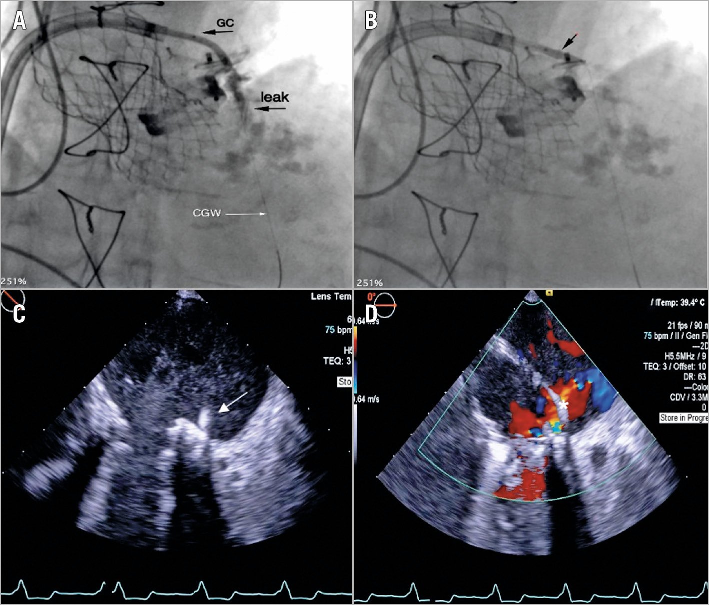
Figure 1. Transseptal PVL closure: procedural steps. A) PVL approach using a 10 Fr steerable catheter. A 6 Fr JR guide catheter is inserted into the 10 Fr catheter; a coronary wire is used for crossing the leak and positioning a 2.5×12 mm Shockwave balloon (black arrow, B). C) TEE shows the Shockwave balloon (white arrow) and microbubbles generated by waves. D) An Occlutech PLD Occluder 8×4 mm (asterisk) is positioned through a 7 Fr catheter.
A 66-year-old female with heart failure was referred for treatment of severe mitral regurgitation (MR) caused by a paravalvular leak (PVL). She underwent four mitral valve replacements (Bicarbon®; Sorin Group/LivaNova, Milan, Italy), three PVL percutaneous closures, and transcatheter aortic valve implantation (CoreValve® 31 mm; Medtronic, Minneapolis, MN, USA). Transoesophageal echocardiography (TEE) showed severe circumferential calcification of the mitral annulus and two PVLs: the first, in an anteromedial position, caused trivial regurgitation; the second one, in a posterolateral position, caused severe MR (Moving image 1). We planned to close the posterolateral PVL because of its haemodynamic significance. A first attempt was unsuccessful because it was impossible to advance any catheter through the calcified leak. A month later, the procedure was performed off-label using an intravascular lithotripsy (IVL) catheter (Shockwave Intravascular Lithotripsy System; Shockwave Medical, Santa Clara, CA, USA). A transseptal puncture was performed in supero-posterior position, followed by septum dilatation (Armada balloon catheter 8.0×40 mm; Abbott Vascular, Santa Clara, CA, USA). A steerable 10 Fr catheter (Destino™; Oscor, Palm Harbor, FL, USA) was positioned in the left atrium (LA) on a stiff wire and used for supporting and orienting a JR4 6 Fr guiding catheter; the leak was crossed with a coronary guidewire (PILOT® 50-0.0014”; Abbott Vascular) (Figure 1A), predilated (NC Euphora™ 2.0×20 mm non-compliant balloon; Medtronic) at 18 atm, to engage the PVL with the Shockwave balloon. A lithotripsy protocol was applied using a Shockwave balloon 2.5×12 mm. In order to avoid annular rupture, the balloon was partially positioned inside the leak and inflated at low pressure (4 mmHg) (Moving image 2, Figure 1B, Figure 1C). The angiogram showed leak modification (Moving image 3) and the PVL was crossed with a JR4 6 Fr catheter and a Terumo wire (Terumo Corp., Somerset, NJ, USA). The system was exchanged over an INNOWI® guidewire (Symedrix, Oberhaching, Germany). A 7 Fr guiding catheter was used to deploy and release an Occlutech® PLD Occluder 8×4 mm waist (Occlutech, Helsingborg, Sweden) (Moving image 4-Moving image 6) with significant reduction of regurgitation (Figure 1D).
IVL was recently successfully applied in the treatment of calcified coronary and peripheral arterial stenosis1. We report the first-in-man IVL application in the treatment of calcified PVL. Further cases will be required to confirm the effectiveness of this technique.
Conflict of interest statement
The authors have no conflicts of interest to declare.
Supplementary data
To read the full content of this article, please download the PDF.
Moving image 1. 3D-TEE moving image of the mitral valve. A view showing the guiding catheter through the posterolateral leak.
Moving image 2. TEE moving image during the application of the Shockwave balloon. A TEE echo view of a Shockwave balloon 2.5×12 mm partially positioned inside the leak. Microbubbles were produced by the application of Shockwave balloon impulses. Three cycles of 10 pulsed waves are applied.
Moving image 3. Intraprocedural angiography after the application of the Shockwave balloon. The angiogram shows a significant enlargement of the leak which allowed easy crossing of the leak with a diagnostic catheter JR4 6 Fr and a Terumo wire.
Moving image 4. Intraprocedural angiography of 7 Fr guiding catheter into a steerable catheter. The fluoroscopy shows the 7 Fr guiding catheter into a 10 Fr steerable catheter (Destino; Oscor), which allowed stability of the system.
Moving image 5. TEE colour image showing the correct deployment of the Occlutech PLD Occluder. The echo moving image confirms the correct position of the device with a significant reduction of paravalvular regurgitation.
Moving image 6. Intraprocedural angiography before the release of the Occlutech PLD Occluder. Final angiographic check of the correct position of the device.