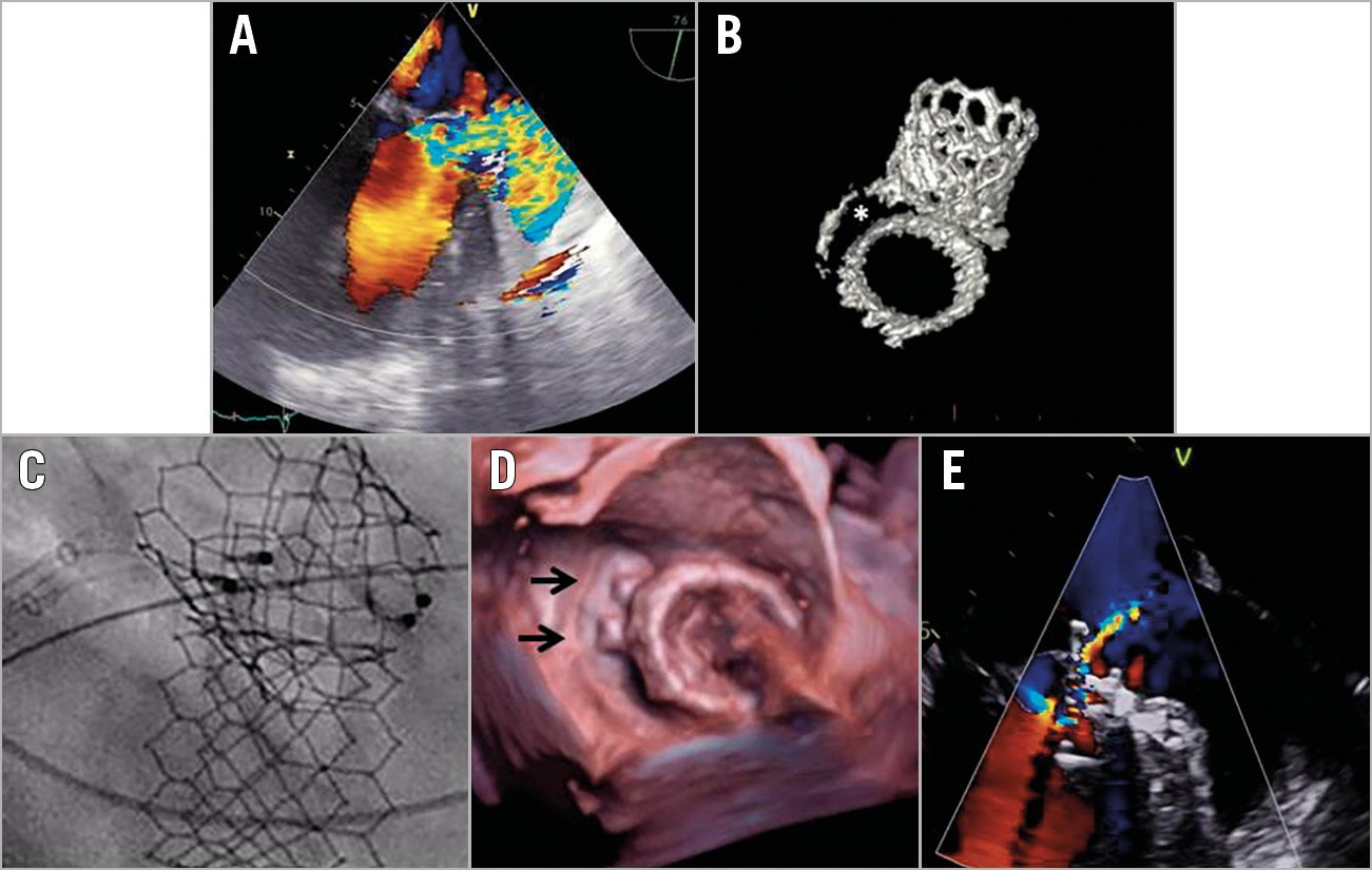
Figure 1. Multimodality imaging of the paravalvular leak and the procedure. A) Transoesophageal echo (TOE) image showing the initial severe paravalvular leak (Moving image 1). B) CT image demonstrating an important anterolateral arch-shaped gap between the annular calcification and the bioprosthesis (indicated by the asterisk). C) The simultaneous positioning of two AMPLATZER Vascular Plug III occluders (5×14 mm) allows an obstruction of the defect between the mitral prosthesis and the calcified mitral annulus (D, occluders are indicated by arrows) along with an important reduction of the regurgitation (E and Moving image 2).
A single-stage transapical aortic then mitral valve implantation was performed in a 68-year-old man. Regarding the mitral valve, an Edwards SAPIEN 3 29 mm bioprosthesis (Edwards Lifesciences, Irvine, CA, USA) was implanted in severe mitral annular calcifications (MAC). Despite initial success, the patient suffered a pulmonary oedema one month after the initial procedure. Echographic and computed tomography analyses revealed a well-functioning aortic prosthesis and a delayed migration of the mitral prosthesis towards the left atrium, resulting in a severe paravalvular leak (PVL) (Figure 1A, Moving image 1, Figure 1B). This PVL was located in the anterolateral part of the mitral valve, with an arch-shaped aspect, measuring 8×16 mm.
A percutaneous (transfemoral transseptal anterograde approach) closure of this PVL was therefore performed under transoesophageal guidance. The simultaneous positioning of two 5×14 mm AMPLATZER™ Vascular Plug (AVP) III occluders (St. Jude Medical [now Abbott Vascular], St. Paul, MN, USA) (Figure 1C, Figure 1D) allowed an important reduction of the PVL (Figure 1E, Moving image 2) without any migration of the mitral prosthesis or left ventricular outflow tract (LVOT) obstruction. At one-month follow-up, the patient was asymptomatic, with a mild residual PVL, and no significant haemolysis. However, at two-month follow-up, his clinical status suddenly worsened, due to a new prosthesis migration: the index PVL correction was still fine, but new ones had appeared. A new 29 mm SAPIEN 3 prosthesis was therefore surgically implanted, with a good result.
Balloon-expandable transcatheter mitral valve implantations in MAC procedures are increasingly performed. Delayed migration of the prosthesis and PVL are well-known complications of these procedures1. However, percutaneous closure of such a PVL can be challenging2. We strongly believe that, because of its oval shape, the AVP III is well suited for such an arch-shaped PVL: positioned in its short axis, it allows a limited protrusion in the LVOT, reducing the risk of obstruction. However, a comprehensive assessment of the underlying mechanism of the PVL remains the cornerstone of any adapted treatment; percutaneous PVL closure should not be performed in case of leak due to valve migration given the risk of further valve embolisation.
Conflict of interest statement
G. Leurent has received fees as speaker/consultant from Abbott, Abiomed, AstraZeneca and Novartis. The other authors have no conflicts of interest to declare.
Supplementary data
To read the full content of this article, please download the PDF.
Moving image 1. Transoesophageal echo (TOE) image showing the initial severe paravalvular leak.
Moving image 2. Residual regurgitation after implantation of two AVP IIIs.