INTERVENTIONAL FLASHLIGHT

DOI: 10.4244/EIJ-D-17-01106

Ad hoc percutaneous paravalvular leak closure after transcatheter aortic valve replacement facilitated by integrated multimodality imaging

Tian-Yuan Xiong1,2, MD; Giuseppe Martucci2, MD; Hind Alosaimi2, MD; Nicolo Piazza2*, MD, PhD

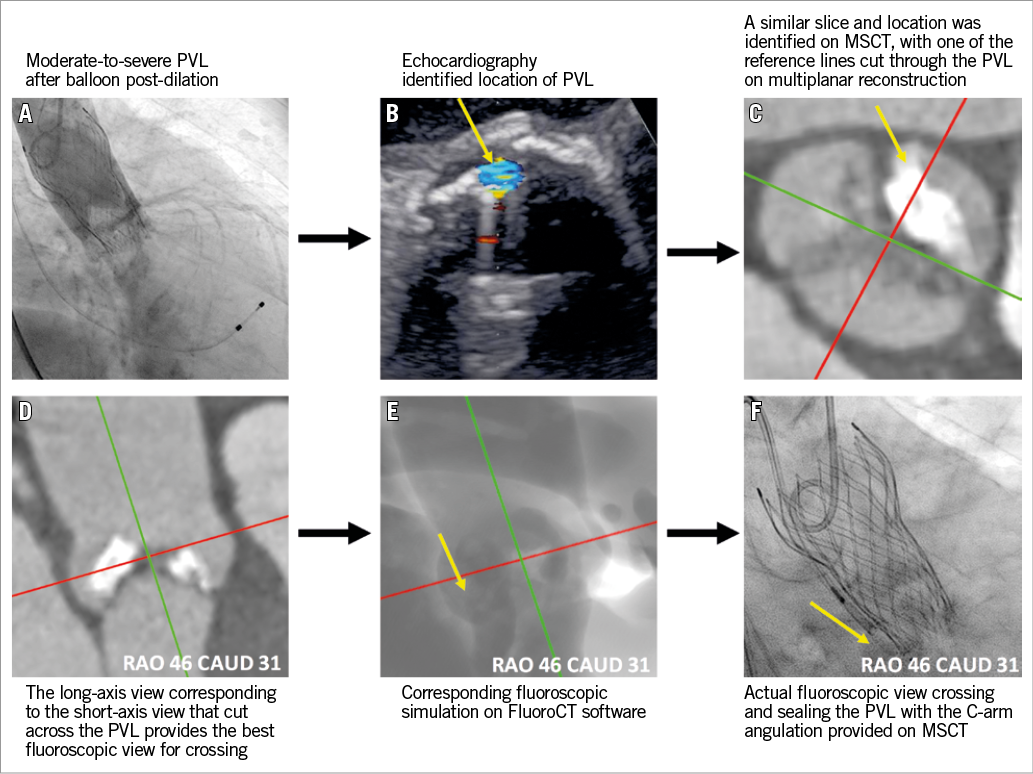
A 91-year-old female with symptomatic severe aortic stenosis (aortic valve area 0.4 cm2, transvalvular mean gradient 77 mmHg) was referred for transcatheter aortic valve replacement (TAVR). A 26 mm CoreValve® Evolut™ R bioprosthesis (Medtronic, Minneapolis, MN, USA) was planned with a perimeter oversizing of 14.6%. A large calcium nodule was noted at the non-coronary cusp on multislice computed tomography (MSCT). Moderate-to-severe paravalvular leak (PVL) was noted on post-deployment aortography. Post-dilation with a 22 mm balloon failed to improve the amount of PVL (Panel A). Considering the significant risk of suboptimal stent apposition and conformability to the surrounding anatomy, even after aggressive post-dilation, and the concern of aortic root perforation/rupture, we chose percutaneous PVL closure to seal the leak.

According to the intraprocedural transoesophageal echocardiogram (TEE), the regurgitant jet was located at the commissure between the left and non-coronary cusps (Panel B). According to the location of the regurgitant jet on the TEE images, we selected an optimal fluoroscopic viewing angle (RAO 46, CAUD 31) for crossing the paravalvular leak after appropriate multiplanar reconstruction of the pre-TAVR MSCT (Panel C-Panel F), using FluoroCT software (Circle Cardiovascular Imaging Inc., Calgary, Canada). The leak was passed with a 0.035-inch J wire, through which an 8 Fr multipurpose guiding catheter crossed the defect. The selected 8 mm AMPLATZER™ Vascular Plug II (St. Jude Medical, St. Paul, MN, USA) was loaded into the delivery system, advanced through the multipurpose guiding catheter and deployed. Position and closure of the leak were confirmed by TEE and angiography. Of note, only 12 minutes were required to analyse the TEE and MSCT images, and cross/plug the PVL.

Percutaneous PVL closure after TAVR is a technically demanding procedure with unpredictable results. Three-dimensional TEE imaging has been shown to benefit such procedures in preprocedural defect sizing, device selection and deployment guidance1. Crossing the PVL defect, however, can still be time-consuming or require exchanges of catheters and guidewires2. The source of these challenges can be difficult anatomies, suboptimal imaging techniques and C-arm angulations. The difficulty in defining the crossing angulation can be easily solved with MSCT while using TEE to identify the location of the defect. Integrated multimodality imaging using echocardiography, MSCT and fluoroscopy was critical to the success of this procedure.

Conflict of interest statement

N. Piazza is a consultant/proctor for HighLife, Medtronic and MicroPort, and is a consultant for Cephea. The other authors have no conflicts of interest to declare.

Volume 14 Number 5
Aug 3, 2018
Volume 14 Number 5
View full issue


Key metrics

On the same subject

Image – Interventional flashlight

10.4244/EIJ-D-19-00744 Apr 17, 2020
Percutaneous closure of paravalvular leak after transcatheter valve implantation in mitral annular calcification
Leurent G et al
free

Image – Interventional flashlight

10.4244/EIJ-D-19-01122 Oct 9, 2020
The retro-antegrade approach to paravalvular leak closure after transcatheter aortic valve replacement
So C et al
free

10.4244/EIJV12SXA11 May 16, 2016
Catheter-based treatment of paravalvular leaks
Taramasso M et al
free

10.4244/EIJV17I9A121 Oct 20, 2021
Transcatheter paravalvular leak closure: catch me if you can
Pilgrim T and Okuno T
free

Image – Interventional flashlight

10.4244/EIJ-D-19-00118 Feb 20, 2020
Feasibility of quantitative assessment of aortic regurgitation in a percutaneous closure of paravalvular leak: expanding videodensitometry
Teixeirense P et al
free

INTERVENTIONAL FLASHLIGHT

10.4244/EIJ-D-17-00647 Jan 19, 2018
Percutaneous closure of subannular rupture following transcatheter aortic valve implantation
Shah S et al
free
Trending articles
338.63

State-of-the-Art Review

10.4244/EIJ-D-21-00904 Apr 1, 2022
Antiplatelet therapy after percutaneous coronary intervention
Angiolillo D et al
free
295.45

Expert consensus

10.4244/EIJ-D-21-00898 Sep 20, 2022
Intravascular ultrasound guidance for lower extremity arterial and venous interventions
Secemsky E et al
free
226.03

State-of-the-Art Review

10.4244/EIJ-D-21-00426 Dec 3, 2021
Myocardial infarction with non-obstructive coronary artery disease
Lindahl B et al
free
209.5

State-of-the-Art Review

10.4244/EIJ-D-21-01034 Jun 3, 2022
Management of in-stent restenosis
Alfonso F et al
free
168.4

Expert review

10.4244/EIJ-D-21-00690 May 15, 2022
Crush techniques for percutaneous coronary intervention of bifurcation lesions
Moroni F et al
free
150.28

State-of-the-Art

10.4244/EIJ-D-22-00776 Apr 3, 2023
Computed tomographic angiography in coronary artery disease
Serruys PW et al
free
103.48

Expert consensus

10.4244/EIJ-E-22-00018 Dec 4, 2023
Definitions and Standardized Endpoints for Treatment of Coronary Bifurcations
Lunardi M et al
free
X

The Official Journal of EuroPCR and the European Association of Percutaneous Cardiovascular Interventions (EAPCI)

EuroPCR EAPCI
PCR ESC
Impact factor: 6.2
2022 Journal Citation Reports®
Science Edition (Clarivate Analytics, 2023)
Online ISSN 1969-6213 - Print ISSN 1774-024X
© 2005-2024 Europa Group - All rights reserved