A 64-year-old patient was admitted with acute lateral wall ST-elevation myocardial infarction. Past medical history was notable for a prior lateral wall myocardial infarction, which was treated with bare metal stent (BMS) implantation in the proximal segment of the LCx. Due to subsequent in-stent restenosis of the BMS as well as a de novo stenosis proximal to the old stent (Moving image 1) a sirolimus-eluting stent (SES, Cypher®; Cordis, Miami Lakes, FL, USA) was implanted nine years ago and covered both lesions.
Later, coronary angiography revealed a known aneurysmal dilation of the coronary vessels with thrombotic occlusion of the proximal LCx in the segment treated with the SES nine years after implantation (Moving image 2). A fresh thrombus was aspirated and optical coherence tomography (OCT) image analysis of the SES stented section showed protruding stent struts, marked saccular evagination and residual thrombus burden (Figure 1A). Histological analysis of the thrombus revealed a massive accumulation of inflammatory cells including neutrophils and mononuclear cells (Figure 1B and Figure 1C).
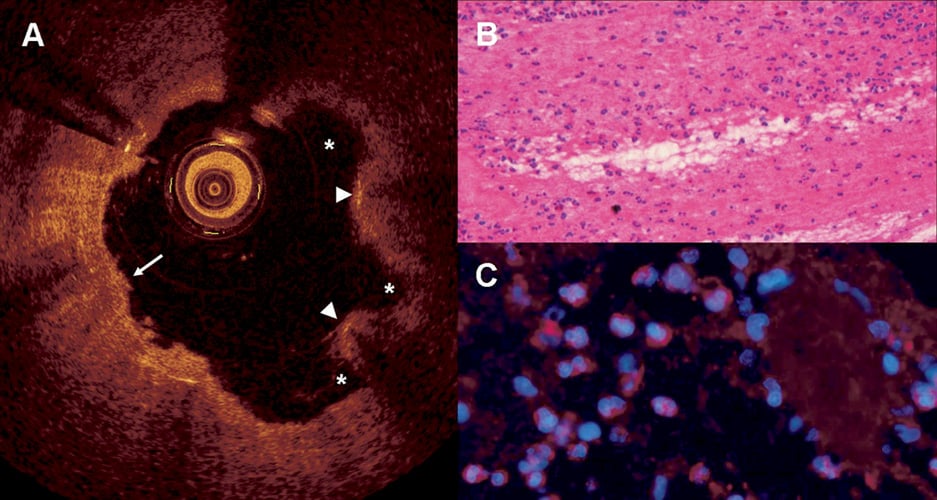
Figure 1. OCT analysis of stent thrombosis and histology of aspirated thrombus. Panel A shows OCT analysis after thrombus aspiration with protruding stent struts (arrowheads) due to substantial saccular evaginations (asterisks) and residual thrombus (arrow). There is some image distortion due to intracatheter blood. Panel B shows massive accumulation of inflammatory cells within the thrombus, in a cross-section following haematoxylin eosin staining, 20x magnification. Panel C shows accumulation of neutrophil granulocytes within the thrombus, following staining of neutrophil elastase in violet and cell nuclei with DAPI (blue), 40x magnification.
Presence of saccular evaginations on OCT imaging probably reflect a pathological cascade of sustained vessel wall inflammation, in response to durable polymer coating, that ultimately resulted in positive remodelling, strut protrusion and very late stent thrombosis.
Funding
PRESTIGE (PREvention of late Stent Thrombosis by an Interdisciplinary Global European effort) project under the Seventh Framework Programme of the European Commission.
Conflict of interest statement
The authors have no conflicts of interest to declare.
Online data supplement
Moving image 1. Projection of left coronary artery with an in-stent restenosis in the LCx as well as a de novo stenosis proximal to the implanted stent.
Moving image 2. Projection of left coronary artery showing acute stent thrombosis of LCx in the proximal segment and known aneurysmal dilation of LAD.