DOI:

Rheolytic versus aspiration thrombectomy in acute st-segment elevation myocardial infarction judged by OCT

Nicolas M. Van Mieghem, MD; Floris Kauer, MD; Evelyn Regar*, MD, PhD

Case description

A 68 year old female presented with an acute ST-segment elevation anterior wall myocardial infarction and was brought to the catheterisation laboratory for a primary percutaneous coronary intervention. After confirmation of a thrombotic occlusion of the left anterior descending coronary artery (LAD) (due to late stent thrombosis) the lesion was crossed with a 0.014” X 180 cm MIRACLEbros 3 guidewire (ASAHI Intecc, Redwood City, CA, USA). The thrombus burden was categorised as TIMI Thrombus Grade 41,2. We proceeded with aspiration thrombectomy using a Thrombuster catheter (Kaneka Medix Corp., Kaneka Corp., Osaka, Japan). The LAD was then evaluated by Optical Coherence Tomography (OCT) with a C7-XR Dragonfly OCT Catheter (LightLab Imaging, Westford, MA, USA) confirming considerable remaining endoluminal thrombus2,3. After introducing a temporary pacemaker into the right ventricle a second run of thombectomy was attempted with the Angiojet Rheolytic thrombectomy catheter (Possis Medical, MN, Minnesota, USA). Control OCT demonstrated an impressive improvement with only minimal residual endoluminal thrombus and pointed towards stent malapposition in the proximal segment (not shown in the image). An 3.5x23 mm XIENCE V DES (Abbott Vascular, Aichi, Japan) was positioned in the proximal LAD. The final angiogram showed brisk TIMI 3 flow with adequate myocardial blush.

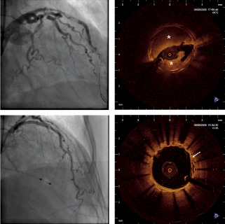
Figure 1. Upper panels: coronary angiogram and OCT image after aspiration thrombectomy: overlapping stent struts and considerable remaining thrombus (*) are clearly visible. Lower panels: coronary angiogram and OCT image after rheolytic thrombectomy with only minimal residual mural thrombus (white arrow).

Volume 5 Number 8
Apr 12, 2010
Volume 5 Number 8
View full issue


Key metrics

Suggested by Cory

Flashlight

10.4244/EIJ-D-24-00989 Apr 7, 2025
Coronary stent retriever-assisted thrombectomy
Walsh J et al
free

Image – Interventional flashlight

10.4244/EIJ-D-20-00985 Aug 27, 2021
Distal embolisation as a cause of ST-segment elevation myocardial infarction: insights from optical coherence tomography
Satogami K et al
free

INTERVENTIONAL FLASHLIGHT

10.4244/EIJ-D-17-00050 Aug 4, 2017
The first reported stentriever-based thrombectomy in acute ST-elevation myocardial infarction due to paradoxical coronary embolism
Manchurov V et al
free

CLINICAL RESEARCH

10.4244/EIJV11I14A315 Apr 8, 2016
Forward and back aspiration during ST-elevation myocardial infarction: a feasibility study
Farooq V et al
free
Trending articles
87.2

State-of-the-Art

10.4244/EIJ-D-25-00266 Jan 19, 2026
Lesion stratification with intracoronary imaging
McGarvey M et al
free
47.45

NEW INNOVATION

10.4244/EIJ-D-15-00467 Feb 20, 2018
Design and principle of operation of the HeartMate PHP (percutaneous heart pump)
Van Mieghem NM et al
free
34.8

Original Research

10.4244/EIJ-D-25-01006 Mar 16, 2026
Clinical outcomes and haemodynamic response after blinded stress assessment of moderate aortic stenosis
Eerdekens R et al
22.2

Viewpoint

10.4244/EIJ-D-25-01066 May 4, 2026
Intracoronary imaging guidance for de novo coronary lesion treatment with drug-coated balloons
Amabile N et al
free
20.75

Flashlight

10.4244/EIJ-D-25-01014 Apr 6, 2026
Stent retriever-assisted coronary thrombectomy with continuous aspiration
Liabot Q et al
open access
19.5

Original Research

10.4244/EIJ-D-26-00032 May 15, 2026
Glucocorticoids to reduce permanent pacemaker implantation after TAVI: the GLUCO-TAVI randomised trial
Fuertes-Kenneally L et al
17.8

Expert Review

10.4244/EIJ-D-25-01316 Apr 20, 2026
Electrosurgical laceration and stabilisation of tricuspid edge-to-edge repair: the ELASTA-T technique
Alvarez-Covarrubias H et al
free
X

PCR
Impact factor: 9.5
2024 Journal Citation Reports®
Science Edition (Clarivate Analytics, 2025)
Online ISSN 1969-6213 - Print ISSN 1774-024X
© 2005-2026 Europa Group - All rights reserved