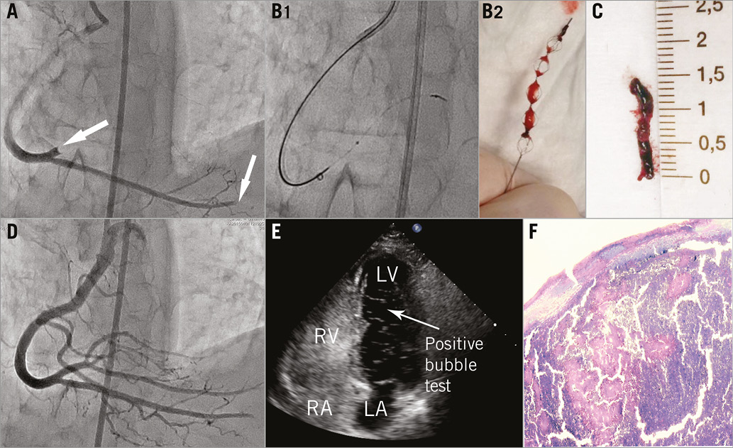

A 54-year-old female was admitted to our hospital suffering from acute inferior ST-elevation myocardial infarction. Emergent coronary angiography showed complete occlusion of both the proximal part of the posterolateral and the distal part of the posterior descending branches of the right coronary artery with no evidence of obstructive coronary atherosclerosis (Panel A, Moving image 1). Angiographic features were strongly suggestive of a coronary embolism. Given the fact that manual thrombus aspiration may be ineffective in cases of coronary embolism and can produce further distal embolisation, we decided to apply stentriever-based thrombectomy. To retrieve the embolus, we used an ERIC® Retrieval Device (Panel B1, Panel B2) with a Sofia™ distal access catheter (both MicroVention, Inc., Tustin, CA, USA) via a 6 Fr Judkins right guiding catheter (Medtronic, Inc., Minneapolis, MN, USA). The technique was applied in the same way1 as that used for thrombectomy in ischaemic stroke (Moving image 2, Moving image 3). The embolus (Panel C) was successfully retrieved and TIMI 3 flow obtained (Panel D, Moving image 4). Post-procedural transthoracic contrast echocardiography revealed the presence of a right-to-left shunt on Valsalva manoeuvre at the atrial septal level (Panel E). Doppler ultrasonography of the lower extremity veins revealed right-sided deep vein thrombosis. Histologic evaluation of the specimen showed signs of organisation and absence of the lines of Zahn, indicating a venous origin of the thrombus (Panel F). The patient’s further clinical course was uneventful. This case shows that stentriever-based thrombectomy might be a safe and feasible approach to treat coronary embolism in selected cases. To our knowledge, this is the first report of the use of a stentriever for the treatment of acute myocardial infarction caused by coronary embolism.
Conflict of interest statement
The authors have no conflicts of interest to declare.
Supplementary data
Moving image 1. Baseline coronary angiogram.
Moving image 2. Stentriever deployment.
Moving image 3. Stentriever pullback.
Moving image 4. Coronary angiogram after thrombectomy.
Supplementary data
To read the full content of this article, please download the PDF.
Baseline coronary angiogram.
Stentriever deployment.
Stentriever pullback.
Coronary angiogram after thrombectomy.

