IMAGE IN CARDIOLOGY

DOI: 10.4244/EIJV12I2A44

The plug for the appendage

Lutz Buellesfeld1*, MD; Stephan Windecker2, MD; Bernhard Meier2, MD

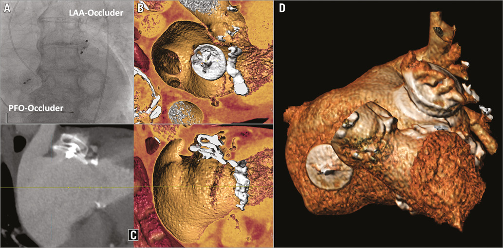
Interventional left atrial appendage (LAA) closure is an emerging treatment for the prevention of atrial clot formation in patients with atrial fibrillation and contraindication for oral anticoagulation. An 82-year-old patient with paroxysmal atrial fibrillation and previous gastrointestinal bleedings underwent implantation of a 28 mm AMPLATZER™ Cardiac Plug (ACP) (St. Jude Medical, St. Paul, MN, USA) within a 24 mm LAA, as well as device closure of a patent foramen ovale grade III (Panel A). The ACP consists of a lobe, which anchors within the atrial appendage, and a disc, which seals the ostium of the LAA (pacifier principle). Multislice computed tomography multiplanar reconstruction and three-dimensional volume rendering demonstrated the successful ACP implantation with optimal disc position one month later (Panel B, Panel C, Moving image 1). However, due to the irregular shape, as well as the thin wall structure of the LAA with a low radial counterforce, implants within the LAA body are able to alter the LAA geometry (Panel D, Moving image 2). This results in inhomogeneous wall stress which may lead to wall injury and subsequent pericardial effusion, which is reported in up to 5% of cases undergoing interventional LAA closure.

Conflict of interest statement

B. Meier receives consulting fees from St. Jude Medical. The other authors have no conflicts of interest to declare.

Supplementary data

Moving image 1. Three-dimensional reconstruction - internal view.

Moving image 2. Three-dimensional reconstruction - external view.

Supplementary data

To read the full content of this article, please download the PDF.

Three-dimensional reconstruction - internal view.

Three-dimensional reconstruction - external view.
Volume 12 Number 2
Jun 10, 2016
Volume 12 Number 2
View full issue


Key metrics

Suggested by Cory

Image – Interventional flashlight

10.4244/EIJ-D-21-00624 Jun 3, 2022
Percutaneous left atrial appendage closure in a surgically ligated left atrial appendage
Wong I et al
free

Image – Interventional flashlight

10.4244/EIJ-D-18-01064 Apr 16, 2019
Management of incomplete surgical left atrial appendage exclusion
Alkhouli M and Raybuck B
free

10.4244/EIJV11I14A307 Apr 8, 2016
Percutaneous left atrial appendage occlusion in 2016
Tzikas A et al
free

CLINICAL RESEARCH

10.4244/EIJY14M07_03 Aug 20, 2015
Double device left atrial appendage closure
Guérios E et al
free

10.4244/EIJV12SXA10 May 16, 2016
Left atrial appendage closure: patient, device and post-procedure drug selection
Tzikas A and Bergmann M
free
Trending articles
151.43

State-of-the-Art

10.4244/EIJ-D-22-00776 Apr 3, 2023
Computed tomographic angiography in coronary artery disease
Serruys PW et al
free
55.9

Clinical research

10.4244/EIJ-D-22-00621 Feb 20, 2023
Long-term changes in coronary physiology after aortic valve replacement
Sabbah M et al
free
54.9

Expert review

10.4244/EIJ-D-21-01010 Jun 24, 2022
Device-related thrombus following left atrial appendage occlusion
Simard T et al
free
43.75

Clinical Research

10.4244/EIJ-D-21-01091 Aug 5, 2022
Lifetime management of patients with symptomatic severe aortic stenosis: a computed tomography simulation study
Medranda G et al
free
39.95

Clinical research

10.4244/EIJ-D-22-00558 Feb 6, 2023
Permanent pacemaker implantation and left bundle branch block with self-expanding valves – a SCOPE 2 subanalysis
Pellegrini C et al
free
X

The Official Journal of EuroPCR and the European Association of Percutaneous Cardiovascular Interventions (EAPCI)

EuroPCR EAPCI
PCR ESC
Impact factor: 7.6
2023 Journal Citation Reports®
Science Edition (Clarivate Analytics, 2024)
Online ISSN 1969-6213 - Print ISSN 1774-024X
© 2005-2024 Europa Group - All rights reserved