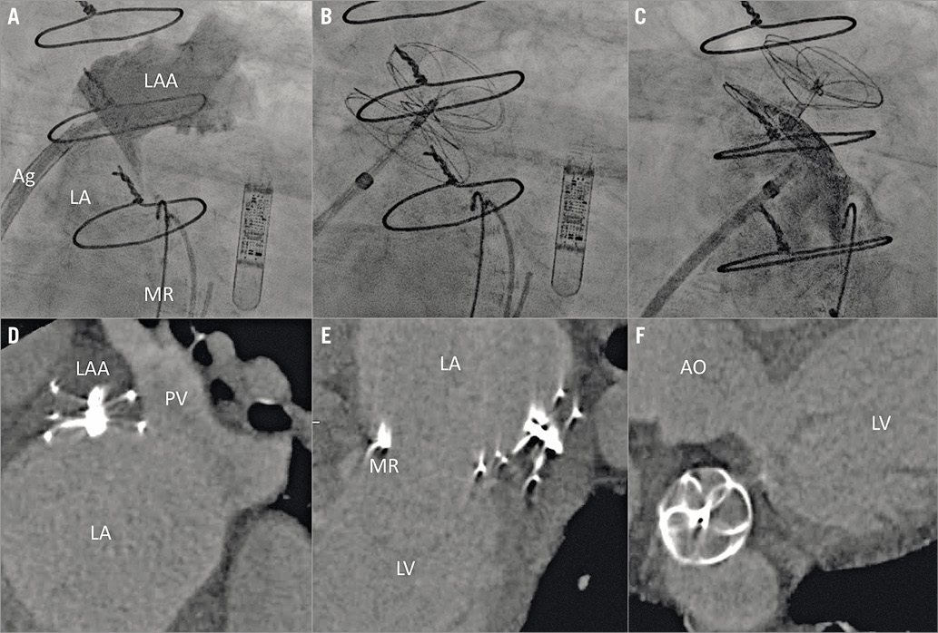
Figure 1. Transcatheter closure of patent left atrial appendage following surgical exclusion. A) LAA angiography via a multipurpose guide. B) A 30 mm Cardioform device deployed across the ostium. C) Left atrial angiography showing complete sealing of the LAA. D) – F) Cardiac computed tomography imaging at one month confirming complete sealing of the LAA. Ag: Agilis sheath; AO: ascending aorta; LA: left atrium; LAA: left atrial appendage; LV: left ventricle; MR: mitral ring; PV: pulmonary vein
A 68-year-old male presented with uncontrolled atrial fibrillation. Medical history included hypertension, arteriovenous malformation, and recent cardiac surgery (mitral and tricuspid annuloplasty, Cox-Maze IV cryoablation, and exclusion of left atrial appendage [LAA]). His CHA2DS2-VASc score was 4 and his HAS-BLED score was 3, but he was not on anticoagulation due to recurrent gastrointestinal bleeding. A transoesophageal echocardiogram performed before cardioversion showed a patent LAA (Moving image 1). Given his intolerance of long-term anticoagulation, he was referred for closure of the LAA.
Transseptal puncture was performed with an Agilis™ sheath (Abbott, Abbott Park, IL, USA), and a Brockenbrough™ needle (Medtronic, Minneapolis, MN, USA). The LAA was cannulated with a 6 Fr multipurpose guide (Medtronic). A pre-curved Amplatz extra-stiff wire (Cook Medical, Bloomington, IN, USA) was then placed in the distal LAA. A 30 mm Cardioform septal occluder (Gore, Flagstaff, AZ, USA) was introduced over the Amplatz wire to the LAA. The left atrial disc was deployed in the LAA and the right atrial disc was deployed in the left atrium bracketing the loosely tied ostium of the LAA (Figure 1A-Figure 1C, Moving image 2, Moving image 3). The patient was discharged home on aspirin 81 mg and Plavix 75 mg. Computed tomography at one month showed complete sealing of the LAA and no device thrombus (Figure 1D-Figure >1F).
Incomplete closure of the LAA after surgical ligation is frequent and is associated with an increased risk of stroke1,2. Transcatheter closure of residual LAA leaks is a feasible and effective management strategy in patients with incomplete surgical LAA closure who are not suitable for long-term anticoagulation.
Conflict of interest statement
The authors have no conflicts of interest to declare.
Supplementary data
Moving image 1. Preprocedural TEE showing a patent LAA.
Moving image 2. Intraprocedural TEE showing percutaneous closure of the LAA.
Moving image 3. Intraprocedural cine imaging illustrating the steps of the closure procedure.
Supplementary data
To read the full content of this article, please download the PDF.
Moving image 1. Preprocedural TEE showing a patent LAA.
Moving image 2. Intraprocedural TEE showing percutaneous closure of the LAA.
Moving image 3. Intraprocedural cine imaging illustrating the steps of the closure procedure.